Human 14-3-3 zeta Antibody Summary
cross-reactivity with recombinant human 14-3-3 beta, theta, eta, gamma, sigma, or epsilon is observed.
Asp2-Asn245
Accession # P63104
Applications
Please Note: Optimal dilutions should be determined by each laboratory for each application. General Protocols are available in the Technical Information section on our website.
Scientific Data
 View Larger
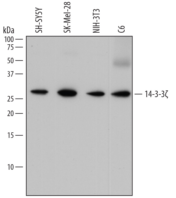
Detection of Human, Mouse, and Rat 14‑3‑3 zeta by Western Blot. Western blot shows lysates of SH-SY5Y human neuroblastoma cell line, SK-Mel-28 human malignant melanoma cell line, NIH-3T3 mouse embryonic fibroblast cell line, and C6 rat glioma cell line. PVDF membrane was probed with 0.2 µg/mL of Mouse Anti-Human 14-3-3 zeta Monoclonal Antibody (Catalog # MAB2669) followed by HRP-conjugated Anti-Mouse IgG Secondary Antibody (Catalog # HAF018). A specific band was detected for 14-3-3 zeta at approximately 27 kDa (as indicated). This experiment was conducted under reducing conditions and using Immunoblot Buffer Group 1.
 View Larger
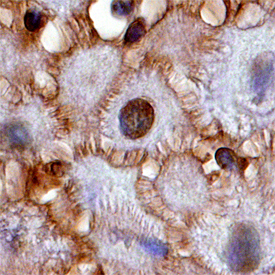
14‑3‑3 zeta in Human Squamous Cell Carcinoma. 14-3-3 zeta was detected in immersion fixed paraffin-embedded sections of human squamous cell carcinoma using Mouse Anti-Human 14-3-3 zeta Monoclonal Antibody (Catalog # MAB2669) at 15 µg/mL overnight at 4 °C. Before incubation with the primary antibody, tissue was subjected to heat-induced epitope retrieval using Antigen Retrieval Reagent-Basic (Catalog # CTS013). Tissue was stained using the Anti-Mouse HRP-DAB Cell & Tissue Staining Kit (brown; Catalog # CTS002) and counter-stained with hematoxylin (blue). Specific staining was localized to nuclei and plasma membrane. View our protocol for Chromogenic IHC Staining of Paraffin-embedded Tissue Sections.
 View Larger
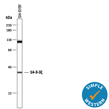
Detection of Human 14‑3‑3 zeta by Simple WesternTM. Simple Western lane view shows lysates of SH‑SY5Y human neuroblastoma cell line, loaded at 0.5 mg/mL. A specific band was detected for 14‑3‑3 zeta at approximately 34 kDa (as indicated) using 2 µg/mL of Mouse Anti-Human 14‑3‑3 zeta Monoclonal Antibody (Catalog # MAB2669). This experiment was conducted under reducing conditions and using the 12-230 kDa separation system.
Reconstitution Calculator
Preparation and Storage
- 12 months from date of receipt, -20 to -70 °C as supplied.
- 1 month, 2 to 8 °C under sterile conditions after reconstitution.
- 6 months, -20 to -70 °C under sterile conditions after reconstitution.
Background: 14-3-3 zeta
14-3-3 proteins are a highly conserved family of homo- and heterodimeric phosphoserine/threonine-binding proteins present in high abundance in all eukaryotic cells. 14-3-3 proteins were the first polypeptides shown to have pSer/Thr binding properties, generally recognizing the consensus sequences RSXpSXP and RXY/FXpSXP (where X is any amino acid). 14-3-3 proteins act as key regulators of intracellular signal transduction through their ability to bind specific motifs phosphorylated on serine or threonine. For example, the binding of 14-3-3 to phosphorylated BAD blocks its proapoptotic association with Bcl-XL. There are at least seven distinct 14-3-3 genes in vertebrates, alpha/beta, epsilon, eta, gamma, theta, sigma and zeta ( alpha / beta, epsilon, eta, gamma, tau / theta, sigma, and zeta /δ). 14-3-3 zeta, also known as Tyrosine 3-Monooxygenase/Tryptophan 5-Monooxygenase Activation Protein, zeta isoform (gene name YWHAZ) is a 245 amino acid, 27 kDa protein.
Product Datasheets
FAQs
No product specific FAQs exist for this product, however you may
View all Antibody FAQsReviews for Human 14-3-3 zeta Antibody
There are currently no reviews for this product. Be the first to review Human 14-3-3 zeta Antibody and earn rewards!
Have you used Human 14-3-3 zeta Antibody?
Submit a review and receive an Amazon gift card.
$25/€18/£15/$25CAN/¥75 Yuan/¥2500 Yen for a review with an image
$10/€7/£6/$10 CAD/¥70 Yuan/¥1110 Yen for a review without an image


