Human ALK-1 Antibody Summary
Asp22-Gln118
Accession # P37023
Applications
Please Note: Optimal dilutions should be determined by each laboratory for each application. General Protocols are available in the Technical Information section on our website.
Scientific Data
 View Larger
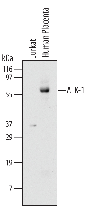
Detection of Human ALK-1 by Western Blot. Western blot shows lysates of Jurkat human acute T cell leukemia cell line and human placenta tissue. PVDF membrane was probed with 2 µg/mL of Mouse Anti-Human ALK-1 Monoclonal Antibody (Catalog # MAB370) followed by HRP-conjugated Anti-Mouse IgG Secondary Antibody (Catalog # HAF007). A specific band was detected for ALK-1 at approximately 65 kDa (as indicated). This experiment was conducted under reducing conditions and using Immunoblot Buffer Group 1.
 View Larger
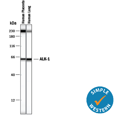
Detection of Human ALK‑1 by Simple WesternTM. Simple Western lane view shows lysates of human placenta tissue and human lung tissue, loaded at 0.2 mg/mL. A specific band was detected for ALK‑1 at approximately 63 kDa (as indicated) using 40 µg/mL of Mouse Anti-Human ALK‑1 Monoclonal Antibody (Catalog # MAB370). This experiment was conducted under reducing conditions and using the 12-230 kDa separation system.Non-specific interaction with the 230 kDa Simple Western standard may be seen with this antibody.
Reconstitution Calculator
Preparation and Storage
- 12 months from date of receipt, -20 to -70 °C as supplied.
- 1 month, 2 to 8 °C under sterile conditions after reconstitution.
- 6 months, -20 to -70 °C under sterile conditions after reconstitution.
Background: ALK-1
Transforming Growth Factor beta (TGF-beta ) superfamily ligands exert their biological activities via binding to heteromeric receptor complexes of two types (I and II) of serine/threonine kinases. Type II receptors are constitutively active kinases that phosphorylate type I receptors upon ligand binding. In turn, activated type I kinases phosphorylate downstream signaling molecules including the various smads. Transmembrane proteoglycans, including the type III receptor (betaglycan) and endoglin, can bind and present some of the TGF-beta superfamily ligands to type I and II receptor complexes and enhance their cellular responses. Seven type I receptors (also termed activin receptor-like kinase (ALK)) and five type II receptors have been isolated from mammals. ALK-2, -3, -4, -5, and -6 are also known as Activin R1A, BMPR-1A, Activin R1B, TGF-beta R1, and BMPR-1B, respectively, reflecting their ligand preferences. Evidence suggests that TGF-beta 1, TGF-beta 3 and an unknown ligand present in serum can activate chimeric ALK-1. ALK-1 shares with other type I receptors a cysteine-rich domain with conserved cysteine spacing in the extracellular region, and a glycine- and serine-rich domain (the GS domain) preceding the kinase domain. ALK-1 is expressed highly in endothelial cells and other highly vascularized tissues. The expression patterns of ALK-1 parallels that of endoglin. Mutations in ALK-1 as well as in endoglin are associated with hereditary hemorrhagic telangiectasia (HHT), suggesting a critical role for ALK-1 in the control of blood vessel development or repair. Human and mouse ALK-1 share approximately 71% amino acid sequence identity in their extracellular regions.
- ten Dijke, P. et al. (1993) Oncogene 8:2879.
- ten Dijke, P. et al. (1994) Science 264:101.
- Lux, A. et al. (1999) J. Biol. Chem. 274:9984.
Product Datasheets
Citations for Human ALK-1 Antibody
R&D Systems personnel manually curate a database that contains references using R&D Systems products. The data collected includes not only links to publications in PubMed, but also provides information about sample types, species, and experimental conditions.
3
Citations: Showing 1 - 3
Filter your results:
Filter by:
-
ALK1 opposes ALK5/Smad3 signaling and expression of extracellular matrix components in human chondrocytes.
Authors: Finnson KW, Parker WL, ten Dijke P, Thorikay M, Philip A
J. Bone Miner. Res., 2008-06-01;23(6):896-906.
Species: Human
Sample Types: Cell Lysates
Applications: Western Blot -
Antitumor activity of ALK1 in pancreatic carcinoma cells.
Authors: Ungefroren H, Schniewind B, Groth S, Chen WB, Muerkoster SS, Kalthoff H, Fandrich F
Int. J. Cancer, 2007-04-15;120(8):1641-51.
Species: Human
Sample Types: Whole Cells
Applications: Western Blot -
Therapeutic action of tranexamic acid in hereditary haemorrhagic telangiectasia (HHT): regulation of ALK-1/endoglin pathway in endothelial cells.
Authors: Fernandez-L A, Garrido-Martin EM, Sanz-Rodriguez F, Ramirez JR, Morales-Angulo C, Zarrabeitia R, Perez-Molino A, Bernabeu C, Botella LM
Thromb. Haemost., 2007-02-01;97(2):254-62.
Species: Human
Sample Types: Whole Cells
Applications: Flow Cytometry
FAQs
No product specific FAQs exist for this product, however you may
View all Antibody FAQsReviews for Human ALK-1 Antibody
There are currently no reviews for this product. Be the first to review Human ALK-1 Antibody and earn rewards!
Have you used Human ALK-1 Antibody?
Submit a review and receive an Amazon gift card.
$25/€18/£15/$25CAN/¥75 Yuan/¥2500 Yen for a review with an image
$10/€7/£6/$10 CAD/¥70 Yuan/¥1110 Yen for a review without an image


