Human Arginase 1/ARG1 PE-conjugated Antibody Summary
Met1-Lys322
Accession # P05089
Applications
Please Note: Optimal dilutions should be determined by each laboratory for each application. General Protocols are available in the Technical Information section on our website.
Scientific Data
 View Larger
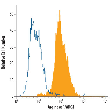
Detection of Arginase 1/ARG1 in HepG2 Human Cell Line by Flow Cytometry. HepG2 human hepatocellular carcinoma cell line was stained with Mouse Anti-Human Arginase 1/ARG1 PE-conjugated Mono-clonal Antibody (Catalog # IC8026P, filled histogram) or isotype control antibody (Catalog # IC0041P, open histogram). To facilitate intracellular staining, cells were fixed with Flow Cytometry Fixation Buffer (Catalog # FC004) and permeabilized with Flow Cytometry Permeabilization/Wash Buffer I (Catalog # FC005). View our protocol for Staining Intracellular Molecules.
 View Larger
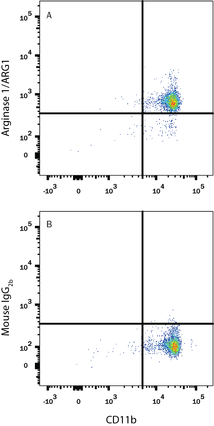
Detection of Arginase 1/ARG1 in Human PBMCs by Flow Cytometry. Human peripheral blood mononuclear cells (PBMCs) were stained with Mouse Anti-Human Integrin aM/CD11b APC-conjugated Monoclonal Antibody (Catalog # FAB1699A) and either (A) Mouse Anti-Human Arginase 1/ARG1 PE-conjugated Monoclonal Antibody (Catalog # IC8026P) or (B) Mouse IgG2BPhycoerythrin Isotype Control (Catalog # IC0041P). To facilitate intracellular staining, cells were fixed with Flow Cytometry Fixation Buffer (Catalog # FC004) and permeabilized with Flow Cytometry Permeabilization/Wash Buffer I (Catalog # FC005). View our protocol for Staining Intracellular Molecules.
Reconstitution Calculator
Preparation and Storage
- 12 months from date of receipt, 2 to 8 °C as supplied.
Background: Arginase 1/ARG1
Arginase 1 (ARG1) is a 35‑40 kDa member of the arginase family of enzymes. It is expressed in multiple cell types, including erythrocytes, hepatocytes, neutrophils, smooth muscle and macrophages. ARG1 demonstrates two distinct functions: in the hepatocyte cytoplasm, it catalyzes the conversion of arginine to ornithine and urea, while in multiple cells, it degrades arginine, thus indirectly downregulating NO synthase (NOS) activity by depriving this enzyme of its substrate. Human ARG1 is 322 amino acids (aa) in length. Its enzyme region comprises aa 9‑309 and contains two Mn atoms. ARG1 is moderately active as a monomer, but highly active as a 105 kDa homotrimer. Trimerization is promoted by nitrosylation of Cys303, creating a regulatory feedback loop with NOS. There are two isoform variants, one that shows an eight aa insertion after Gln43, and another that shows a deletion of aa 204‑289. Full-length human ARG1 shares 87% aa identity with mouse and rat ARG1.
Product Datasheets
Citations for Human Arginase 1/ARG1 PE-conjugated Antibody
R&D Systems personnel manually curate a database that contains references using R&D Systems products. The data collected includes not only links to publications in PubMed, but also provides information about sample types, species, and experimental conditions.
5
Citations: Showing 1 - 5
Filter your results:
Filter by:
-
Metabolic reprogramming of tumor-associated macrophages by collagen turnover promotes fibrosis in pancreatic cancer
Authors: MM LaRue, S Parker, J Puccini, M Cammer, AC Kimmelman, D Bar-Sagi
Proceedings of the National Academy of Sciences of the United States of America, 2022-04-11;119(16):e2119168119.
Species: Mouse
Sample Types: Whole Cells
Applications: Flow Cytometry -
Distinct Populations of Immune-Suppressive Macrophages Differentiate from Monocytic Myeloid-Derived Suppressor Cells in Cancer
Authors: T Kwak, F Wang, H Deng, T Condamine, V Kumar, M Perego, A Kossenkov, LJ Montaner, X Xu, W Xu, C Zheng, LM Schuchter, RK Amaravadi, TC Mitchell, GC Karakousis, C Mulligan, B Nam, G Masters, N Hockstein, J Bennett, Y Nefedova, DI Gabrilovic
Cell Reports, 2020-12-29;33(13):108571.
Species: Human
Sample Types: Whole Cells
Applications: Flow Cytometry -
High M-MDSC Percentage as a Negative Prognostic Factor in Chronic Lymphocytic Leukaemia
Authors: M Zarobkiewi, W Kowalska, S Chocholska, W Tomczak, A Szyma?ska, I Morawska, A Wojciechow, A Bojarska-J
Cancers (Basel), 2020-09-14;12(9):.
Species: Human
Sample Types: Whole Cells
Applications: ICC -
Monocyte Differentiation towards Protumor Activity Does Not Correlate with M1 or M2 Phenotypes
Authors: G Karina Chimal-Ram
J Immunol Res, 2016-06-08;2016(0):6031486.
Species: Human
Sample Types: Whole Cells
Applications: Flow Cytometry -
Ubiquitous Over-Expression of Chromatin Remodeling Factor SRG3 Ameliorates the T Cell-Mediated Exacerbation of EAE by Modulating the Phenotypes of both Dendritic Cells and Macrophages.
Authors: Lee S, Park H, Jeon S, Lee C, Seong R, Park S, Hong S
PLoS ONE, 2015-07-06;10(7):e0132329.
Species: Human
Sample Types: Whole Cells
Applications: Flow Cytometry
FAQs
No product specific FAQs exist for this product, however you may
View all Antibody FAQsReviews for Human Arginase 1/ARG1 PE-conjugated Antibody
Average Rating: 5 (Based on 1 Review)
Have you used Human Arginase 1/ARG1 PE-conjugated Antibody?
Submit a review and receive an Amazon gift card.
$25/€18/£15/$25CAN/¥75 Yuan/¥2500 Yen for a review with an image
$10/€7/£6/$10 CAD/¥70 Yuan/¥1110 Yen for a review without an image
Filter by:
I used this antibody to stain human neutrophils from blood and synovial fluid of arthritis patients. Blood neutrophils had low surface expression of arginase 1, while neutrophils isolated from the synovial fluid of the inflamed knee were found to retain this enzyme on the cell surface. This antibody may be used to stain other cell types as well including macrophages. Cells were stained on ice to minimize internalization of the antibody, but I did not assess cross-reactivity with the mitochondria-association isoform arginase-2.


