Human/Canine Integrin beta 1/CD29 Antibody Summary
Gln21-Asp728
Accession # P05556
Applications
Please Note: Optimal dilutions should be determined by each laboratory for each application. General Protocols are available in the Technical Information section on our website.
Scientific Data
 View Larger
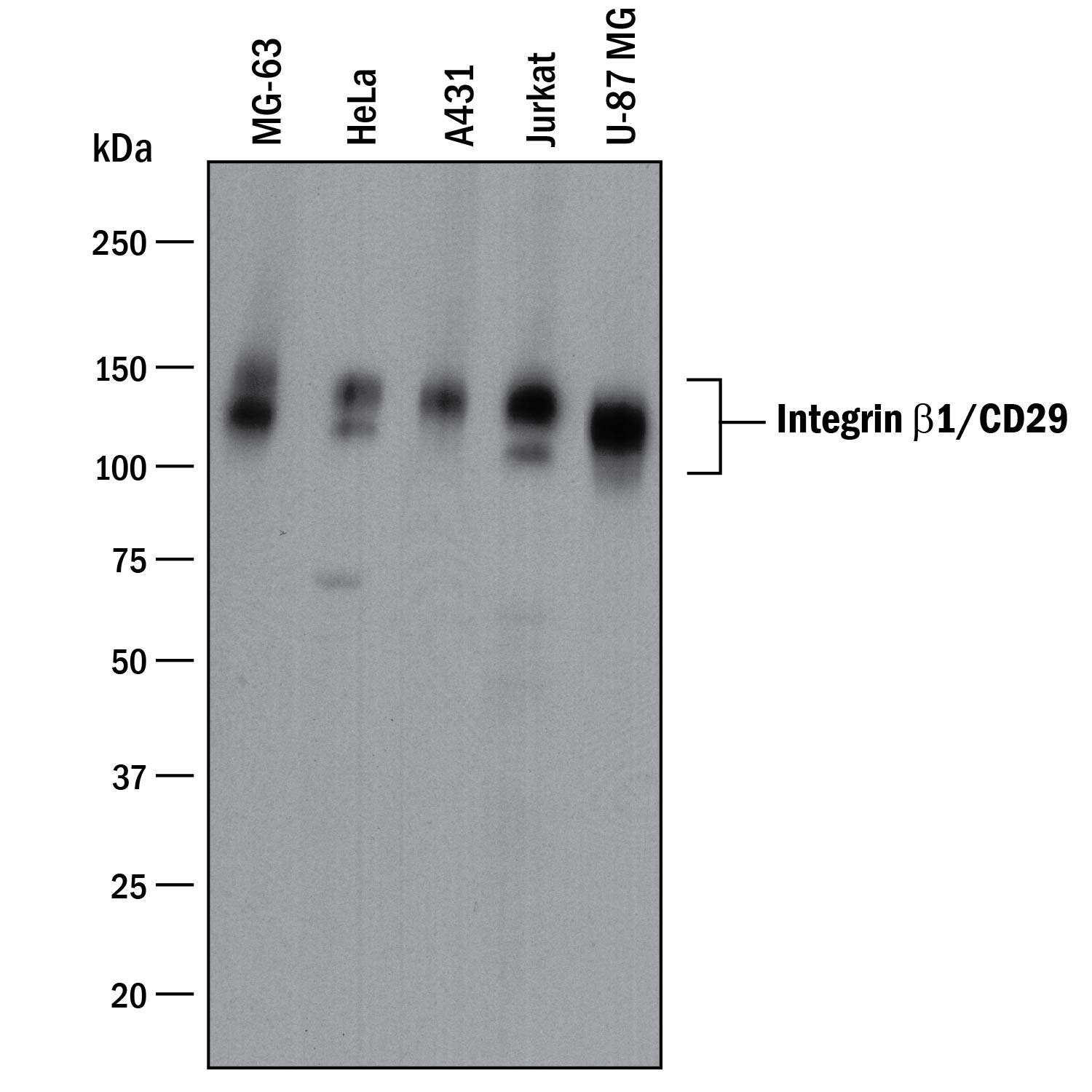
Detection of Human Integrin beta 1/CD29 by Western Blot. Western blot shows lysates of MG-63 human osteosarcoma cell line, HeLa human cervical epithelial carcinoma cell line, A431 human epithelial carcinoma cell line, Jurkat human acute T cell leukemia cell line, and U-87 MG human glioblastoma/astrocytoma cell line. PVDF membrane was probed with 1 µg/mL of Goat Anti-Human/Canine Integrin beta 1/CD29 Antigen Affinity-purified Polyclonal Antibody (Catalog # AF1778) followed by HRP-conjugated Anti-Goat IgG Secondary Antibody (Catalog # HAF017). Specific bands were detected for Integrin beta 1/CD29 at approximately 130-140 kDa (as indicated). This experiment was conducted under reducing conditions and using Immunoblot Buffer Group 1.
 View Larger
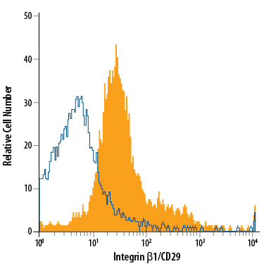
Detection of Integrin beta 1/CD29 in Canine PBMCs by Flow Cytometry. Canine peripheral blood mononuclear cells (PBMCs) were stained with Goat Anti-Human/Canine Integrin beta 1/CD29 Antigen Affinity-purified Polyclonal Antibody (Catalog # AF1778, filled histogram) or isotype control antibody (Catalog # AB-108-C, open histogram), followed by Phycoerythrin-conjugated Anti-Goat IgG Secondary Antibody (Catalog # F0107).
 View Larger
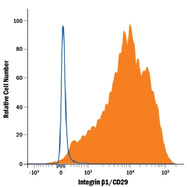
Detection of Integrin beta 1/CD29 in Human PBMCs by Flow Cytometry. Human peripheral blood mononuclear cells (PBMCs) were stained with Goat Anti-Human/Canine Integrin beta 1/CD29 Antigen Affinity-purified Polyclonal Antibody (Catalog # AF1778, filled histogram) or isotype control antibody (Catalog # AB-108-C, open histogram), followed by Phycoerythrin-conjugated Anti-Goat IgG Secondary Antibody (Catalog # F0107).
 View Larger
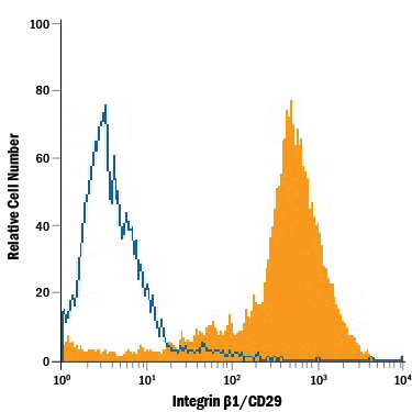
Detection of Integrin beta 1/CD29 in Canine Mesenchymal Stem Cells by Flow Cytometry. Canine mesenchymal stem cells were stained with Goat Anti-Human/Canine Integrin beta 1/CD29 Antigen Affinity-purified Polyclonal Antibody (Catalog # AF1778, filled histogram) or isotype control antibody (Catalog # AB-108-C, open histogram), followed by Phycoerythrin-conjugated Anti-Goat IgG Secondary Antibody (Catalog # F0107).
 View Larger
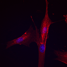
Integrin beta 1/CD29 in Canine Mesenchymal Stem Cells. Integrin beta 1/CD29 was detected in immersion fixed canine mesenchymal stem cells using Goat Anti-Human/Canine Integrin beta 1/CD29 Antigen Affinity-purified Polyclonal Antibody (Catalog # AF1778) at 10 µg/mL for 3 hours at room temperature. Cells were stained using the NorthernLights™ 557-conjugated Anti-Goat IgG Secondary Antibody (red; Catalog # NL001) and counterstained with DAPI (blue). Specific staining was localized to cell surfaces. View our protocol for Fluorescent ICC Staining of Stem Cells on Coverslips.
 View Larger
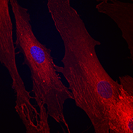
Integrin beta 1/CD29 in Human Mesenchymal Stem Cells. Integrin beta 1/CD29 was detected in immersion fixed human mesenchymal stem cells using Goat Anti-Human/Canine Integrin beta 1/CD29 Antigen Affinity-purified Polyclonal Antibody (Catalog # AF1778) at 10 µg/mL for 3 hours at room temperature. Cells were stained using the NorthernLights™ 557-conjugated Anti-Goat IgG Secondary Antibody (red; Catalog # NL001) and counterstained with DAPI (blue). Specific staining was localized to cell surfaces. View our protocol for Fluorescent ICC Staining of Stem Cells on Coverslips.
 View Larger
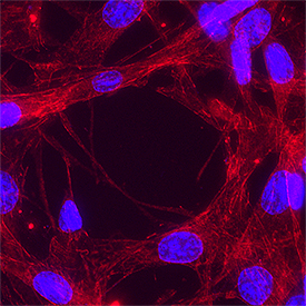
Integrin beta 1/CD29 in A549 Human Cell Line. Integrin beta 1/CD29 was detected in immersion fixed A549 human lung carcinoma cell line using Goat Anti-Human/Canine Integrin beta 1/CD29 Antigen Affinity-purified Polyclonal Antibody (Catalog # AF1778) at 5 µg/mL for 3 hours at room temperature. Cells were stained using the NorthernLights™ 557-conjugated Anti-Goat IgG Secondary Antibody (red; NL001) and counterstained with DAPI (blue). Specific staining was localized to cytoplasm. Staining was performed using our protocol for Fluorescent ICC Staining of Non-adherent Cells.
 View Larger
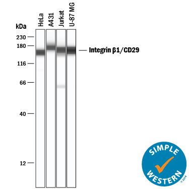
Detection of Human Integrin beta 1/CD29 by Simple WesternTM. Simple Western lane view shows lysates of HeLa human cervical epithelial carcinoma cell line, A431 human epithelial carcinoma cell line, Jurkat human acute T cell leukemia cell line, and U-87 MG human glioblastoma/astrocytoma cell line, loaded at 0.2 mg/mL. Specific bands were detected for Integrin beta 1/CD29 at approximately 157-176 kDa (as indicated) using 10 µg/mL of Goat Anti-Human/Canine Integrin beta 1/CD29 Antigen Affinity-purified Polyclonal Antibody (Catalog # AF1778) followed by 1:50 dilution of HRP-conjugated Anti-Goat IgG Secondary Antibody (Catalog # HAF109). This experiment was conducted under reducing conditions and using the 12-230 kDa separation system.
 View Larger
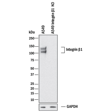
Western Blot Shows Human Integrin beta 1/CD29 Specificity by Using Knockout Cell Line. Western blot shows lysates of A549 human lung carcinoma parental cell line and Integrin beta 1/CD29 knockout A549 cell line (KO). PVDF membrane was probed with 1 µg/mL of Goat Anti-Human/Canine Integrin beta 1/CD29 Antigen Affinity-purified Polyclonal Antibody (Catalog # AF1778) followed by HRP-conjugated Anti-Goat IgG Secondary Antibody (Catalog # HAF017). Specific bands were detected for Integrin beta 1/CD29 at approximately 105 kDa and 120 kDa (as indicated) in the parental A549 cell line, but is not detectable in knockout A549 cell line. GAPDH (Catalog # MAB5718) is shown as a loading control. This experiment was conducted under reducing conditions and using Immunoblot Buffer Group 1.
Reconstitution Calculator
Preparation and Storage
- 12 months from date of receipt, -20 to -70 °C as supplied.
- 1 month, 2 to 8 °C under sterile conditions after reconstitution.
- 6 months, -20 to -70 °C under sterile conditions after reconstitution.
Background: Integrin beta 1/CD29
Integrin beta 1, also called CD29 and VLA-beta chain, associates with at least ten different integrin alpha subunits to form various VLA complexes. The beta 1 subunit has a broad tissue distribution except erythrocytes. Over aa 21-720, human Integrin beta 1 shares 95% aa identity with canine Integrin beta 1.
Product Datasheets
Citations for Human/Canine Integrin beta 1/CD29 Antibody
R&D Systems personnel manually curate a database that contains references using R&D Systems products. The data collected includes not only links to publications in PubMed, but also provides information about sample types, species, and experimental conditions.
3
Citations: Showing 1 - 3
Filter your results:
Filter by:
-
Reparative effect of mesenchymal stromal cells on endothelial cells after hypoxic and inflammatory injury
Authors: JM Sierra-Par, A Merino, M Eijken, H Leuvenink, R Ploeg, BK Møller, B Jespersen, CC Baan, MJ Hoogduijn
Stem Cell Res Ther, 2020-08-12;11(1):352.
Species: Human
Sample Types: Whole Cells
Applications: Neutralization -
Bystander cells enhance NK cytotoxic efficiency by reducing search time
Authors: X Zhou, R Zhao, K Schwarz, M Mangeat, EC Schwarz, M Hamed, I Bogeski, V Helms, H Rieger, B Qu
Sci Rep, 2017-03-13;7(0):44357.
Species: Human
Sample Types: Whole Cells
Applications: Migration Assay -
In vivo biomarker expression patterns are preserved in 3D cultures of Prostate Cancer.
Authors: Windus L, Kiss D, Glover T, Avery V
Exp Cell Res, 2012-07-27;318(19):2507-19.
Species: Human
Sample Types: Cell Lysates
Applications: Western Blot
FAQs
No product specific FAQs exist for this product, however you may
View all Antibody FAQsReviews for Human/Canine Integrin beta 1/CD29 Antibody
There are currently no reviews for this product. Be the first to review Human/Canine Integrin beta 1/CD29 Antibody and earn rewards!
Have you used Human/Canine Integrin beta 1/CD29 Antibody?
Submit a review and receive an Amazon gift card.
$25/€18/£15/$25CAN/¥75 Yuan/¥2500 Yen for a review with an image
$10/€7/£6/$10 CAD/¥70 Yuan/¥1110 Yen for a review without an image


