Human Cathepsin B Antibody Summary
Arg18-Ile339
Accession # P07858
Applications
Please Note: Optimal dilutions should be determined by each laboratory for each application. General Protocols are available in the Technical Information section on our website.
Scientific Data
 View Larger
Detection of Human Cathepsin B by Western Blot. Western blot shows lysates of HepG2 human hepatocellular carcinoma cell line. PVDF membrane was probed with 0.25 µg/mL of Goat Anti-Human Cathepsin B Antigen Affinity-purified Polyclonal Antibody (Catalog # AF953) followed by HRP-conjugated Anti-Goat IgG Secondary Antibody (Catalog # HAF019). A specific band was detected for Cathepsin B at approximately 25-30 kDa (as indicated). This experiment was conducted under reducing conditions and using Immunoblot Buffer Group 1.
.") View Larger
View Larger
Cathepsin B in Human Brain. Cathepsin B was detected in immersion fixed paraffin-embedded sections of human brain (cortex) using Goat Anti-Human Cathepsin B Antigen Affinity-purified Polyclonal Antibody (Catalog # AF953) at 10 µg/mL overnight at 4 °C. Tissue was stained using the Anti-Goat HRP-DAB Cell & Tissue Staining Kit (brown; CTS008) and counterstained with hematoxylin (blue). Lower panel shows a lack of labeling if primary antibodies are omitted and tissue is stained only with secondary antibody followed by incubation with detection reagents. View our protocol for Chromogenic IHC Staining of Paraffin-embedded Tissue Sections.
 View Larger
Detection of Human Cathepsin B by Simple WesternTM. Simple Western lane view shows lysates of HepG2 human hepatocellular carcinoma cell line, loaded at 0.2 mg/mL. A specific band was detected for Cathepsin B at approximately 34 kDa (as indicated) using 2.5 µg/mL of Goat Anti-Human Cathepsin B Antigen Affinity-purified Polyclonal Antibody (Catalog # AF953) followed by 1:50 dilution of HRP-conjugated Anti-Goat IgG Secondary Antibody (HAF109). This experiment was conducted under reducing conditions and using the 12-230 kDa separation system.
 View Larger
Western Blot Shows Human Cathepsin B Specificity by Using Knockout Cell Line. Western blot shows lysates of HeLa human cervical epithelial carcinoma parental cell line and Cathepsin B knockout HeLa cell line (KO). PVDF membrane was probed with 0.25 µg/mL of Goat Anti-Human Cathepsin B Antigen Affinity-purified Polyclonal Antibody (Catalog # AF953) followed by HRP-conjugated Anti-Goat IgG Secondary Antibody (HAF017). Specific bands were detected for Cathepsin B at approximately 25-45 kDa (as indicated) in the parental HeLa cell line, but is not detectable in knockout HeLa cell line. GAPDH (Catalog # AF5718) is shown as a loading control. This experiment was conducted under reducing conditions and using Immunoblot Buffer Group 1. New adjunct appears with knockout cell line.
 View Larger
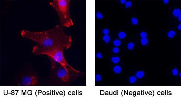
Detection of Cathepsin B in U‑87 (Positive) & Daudi (Negative). Cathepsin B was detected in immersion fixed U‑87 MG human glioblastoma/astrocytoma cell line (Positive) & absent in Daudi human Burkitt's lymphoma cell line (Negative) using Goat Anti-Human Cathepsin B Antigen Affinity-purified Polyclonal Antibody (Catalog # AF953) at 5 µg/mL for 3 hours at room temperature. Cells were stained using the NorthernLights™ 557-conjugated Anti-Goat IgG Secondary Antibody (red; Catalog # NL001) and counterstained with DAPI (blue). Specific staining was localized to cell surface. View our protocol for Fluorescent ICC Staining of Cells on Coverslips.
Reconstitution Calculator
Preparation and Storage
- 12 months from date of receipt, -20 to -70 °C as supplied.
- 1 month, 2 to 8 °C under sterile conditions after reconstitution.
- 6 months, -20 to -70 °C under sterile conditions after reconstitution.
Background: Cathepsin B
Cathepsin B is the first described member of the family of lysosomal cysteine proteases (1). Cathepsin B possesses both endopeptidase and exopeptidase activities, in the latter case acting as a peptidyl-dipeptidase. It is known to process a number of proteins, including pro and active caspases, prorenin, and secretory leucoprotease inhibitor (SLPI) (2-4). Therefore, Cathepsin B may play a role in activation and inactivation of caspases, activation of renin and inactivation of SLPI, the key steps in apoptosis, angiotensin production, and progression of emphysema, respectively. Because of its increased levels and redistribution of the enzyme in human and animal tumors, Cathepsin B may also have role in invasion and metastasis (5).
In addition to lysosome, Cathepsin B can be secreted or associated with plasma membrane, cytoplasm, and nucleus. It is synthesized as a preproenzyme. Following removal of the signal peptide, the inactive proenzyme undergoes further modifications including removal of the pro region to result in the active enzyme (1).
- Mort, J.S. (2004) in Handbook of Proteolytic Enzymes. Barrett, A.J. et al. (eds): Academic Press, San Diego, p. 1079.
- Vancompernolle, K. et al. (1998) FEBS Lett. 438:150.
- Jutras, I. and T.L. Reudelhuber (1999) FEBS Lett. 443:48.
- Taggart, C.C. et al. (2001) J. Biol. Chem. 276:33345.
- Bergquin, I.M. and B.F. Sloane (1996) Adv. Exp. Med. Biol. 389:281.
Product Datasheets
Citations for Human Cathepsin B Antibody
R&D Systems personnel manually curate a database that contains references using R&D Systems products. The data collected includes not only links to publications in PubMed, but also provides information about sample types, species, and experimental conditions.
21
Citations: Showing 1 - 10
Filter your results:
Filter by:
-
Reciprocal effects of alpha-synuclein aggregation and lysosomal homeostasis in synucleinopathy models
Authors: Drobny A, Boros FA, Balta D et al.
Translational neurodegeneration
-
A Practical and Analytical Comparative Study of Gel-Based Top-Down and Gel-Free Bottom-Up Proteomics Including Unbiased Proteoform Detection
Authors: Ercan H, Resch U, Hsu F et al.
Cells
-
Salivary gland LAMP3 mRNA expression is a possible predictive marker in the response to hydroxychloroquine in Sj�gren's disease
Authors: H Nakamura, T Tanaka, Y Ji, C Zheng, SA Afione, BM Warner, FR Oliveira, ACF Motta, EM Rocha, M Noguchi, T Atsumi, JA Chiorini
PLoS ONE, 2023-02-23;18(2):e0282227.
Species: Human
Sample Types: Cell Lysates
Applications: Western Blot -
An infection-induced oxidation site regulates legumain processing and tumor growth
Authors: Y Kovalyova, DW Bak, EM Gordon, C Fung, JHB Shuman, TL Cover, MR Amieva, E Weerapana, SK Hatzios
Nature Chemical Biology, 2022-03-24;0(0):.
Species: Gerbil
Sample Types: Cell Lysates
Applications: Western Blot -
Impact of DJ-1 and Helix 8 on the Proteome and Degradome of Neuron-Like Cells
Authors: U Kern, K Fröhlich, J Bedacht, N Schmidt, ML Biniossek, N Gensch, K Baerenfall, O Schilling
Cells, 2021-02-16;10(2):.
Species: Human
Sample Types: Cell Lysates
Applications: Western Blot -
Cathepsin Inhibition Modulates Metabolism and Polarization of Tumor-Associated Macrophages
Authors: D Oelschlaeg, T Weiss Sada, S Salpeter, S Krug, G Blum, W Schmitz, A Schulze, P Michl
Cancers (Basel), 2020-09-10;12(9):.
Species: Human
Sample Types: Whole Tissue
Applications: IHC -
YWHA/14-3-3 proteins recognize phosphorylated TFEB by a noncanonical mode for controlling TFEB cytoplasmic localization
Authors: Y Xu, J Ren, X He, H Chen, T Wei, W Feng
Autophagy, 2019-01-27;0(0):1-14.
Species: Human
Sample Types: Cell Lysates
Applications: Western Blot -
Modulation of Receptor Protein Tyrosine Phosphatase Sigma Increases Chondroitin Sulfate Proteoglycan Degradation through Cathepsin B Secretion to Enhance Axon Outgrowth
Authors: AP Tran, S Sundar, M Yu, BT Lang, J Silver
J. Neurosci., 2018-05-14;38(23):5399-5414.
Species: Rat
Sample Types: Cell Culture Supernates, Whole Cells, Whole Tissue
Applications: ICC, IHC, Western Blot -
The lysosomal protein cathepsin L is a progranulin protease
Authors: CW Lee, JN Stankowski, J Chew, CN Cook, YW Lam, S Almeida, Y Carlomagno, KF Lau, M Prudencio, FB Gao, M Bogyo, DW Dickson, L Petrucelli
Mol Neurodegener, 2017-07-25;12(1):55.
Species: Human
Sample Types: Cell Lysates
Applications: Western Blot -
The Unusual Resistance of Avian Defensin AvBD7 to Proteolytic Enzymes Preserves Its Antibacterial Activity
PLoS ONE, 2016-08-25;11(8):e0161573.
Species: Chicken
Sample Types: Tissue Homogenates
Applications: Western Blot -
Cathepsin S attenuates endosomal EGFR signalling: A mechanical rationale for the combination of cathepsin S and EGFR tyrosine kinase inhibitors
Sci Rep, 2016-07-08;6(0):29256.
Species: Human
Sample Types: Cell Lysates
Applications: Western Blot -
Simvastatin inhibits glucose metabolism and legumain activity in human myotubes.
Authors: Smith R, Solberg R, Jacobsen L, Voreland A, Rustan A, Thoresen G, Johansen H
PLoS ONE, 2014-01-08;9(1):e85721.
Species: Human
Sample Types: Cell Lysates, Whole Cells
Applications: ICC, Western Blot -
Human bone marrow-derived mesenchymal stem cells suppress human glioma growth through inhibition of angiogenesis.
Authors: Ho I, Toh H, Ng W, Teo Y, Guo C, Hui K, Lam P
Stem Cells, 2013-01-01;31(1):146-55.
Species: Human
Sample Types: Whole Tissue
Applications: IHC-Fr -
Differential secretome analysis reveals CST6 as a suppressor of breast cancer bone metastasis.
Cell Res., 2012-06-12;22(9):1356-73.
Species: Human
Sample Types: Cell Lysates
Applications: Western Blot -
A possible contribution of altered cathepsin B expression to the development of skin sclerosis and vasculopathy in systemic sclerosis.
Authors: Noda S, Asano Y, Akamata K, Aozasa N, Taniguchi T, Takahashi T, Ichimura Y, Toyama T, Sumida H, Yanaba K, Tada Y, Sugaya M, Kadono T, Sato S
PLoS ONE, 2012-02-23;7(2):e32272.
Species: Human
Sample Types: Whole Tissue
Applications: IHC-P -
Macrophages and cathepsin proteases blunt chemotherapeutic response in breast cancer.
Authors: Shree T, Olson OC, Elie BT
Genes Dev., 2011-12-01;25(23):2465-79.
Species: Human
Sample Types: Whole Tissue
Applications: IHC-P -
CD40 stimulation sensitizes CLL cells to lysosomal cell death induction by type II anti-CD20 mAb GA101.
Authors: Jak M, van Bochove GG, Reits EA
Blood, 2011-09-26;118(19):5178-88.
Species: Human
Sample Types: Cell Lysates
Applications: Western Blot -
Transgenic expression of human cathepsin B promotes progression and metastasis of polyoma-middle-T-induced breast cancer in mice.
Authors: Sevenich L, Werner F, Gajda M, Schurigt U, Sieber C, Muller S, Follo M, Peters C, Reinheckel T
Oncogene, 2010-09-06;30(1):54-64.
Species: Human
Sample Types: Tissue Homogenates, Whole Tissue
Applications: IHC-P, Western Blot -
The NALP3 inflammasome is involved in the innate immune response to amyloid-beta.
Authors: Halle A, Hornung V, Petzold GC, Stewart CR, Monks BG, Reinheckel T, Fitzgerald KA, Latz E, Moore KJ, Golenbock DT
Nat. Immunol., 2008-07-11;9(8):857-65.
Species: Mouse
Sample Types: Whole Cells
Applications: ICC -
Distinct roles for cysteine cathepsin genes in multistage tumorigenesis.
Authors: Gocheva V, Zeng W, Ke D, Klimstra D, Reinheckel T, Peters C, Hanahan D, Joyce JA
Genes Dev., 2006-02-15;20(5):543-56.
Species: Human
Sample Types: Whole Tissue
Applications: IHC-P -
Biomarker discovery from pancreatic cancer secretome using a differential proteomic approach.
Authors: Gronborg M, Kristiansen TZ, Iwahori A, Chang R, Reddy R, Sato N, Molina H, Jensen ON, Hruban RH, Goggins MG, Maitra A, Pandey A
Mol. Cell Proteomics, 2005-10-08;5(1):157-71.
Species: Human
Sample Types: Cell Lysates
Applications: Western Blot
FAQs
No product specific FAQs exist for this product, however you may
View all Antibody FAQsReviews for Human Cathepsin B Antibody
Average Rating: 5 (Based on 3 Reviews)
Have you used Human Cathepsin B Antibody?
Submit a review and receive an Amazon gift card.
$25/€18/£15/$25CAN/¥75 Yuan/¥2500 Yen for a review with an image
$10/€7/£6/$10 CAD/¥70 Yuan/¥1110 Yen for a review without an image
Filter by:
1:500 dilution is good.

It's better to use 12% SDS-PAGE


