Human Cathepsin D Antibody Summary
Leu21-Leu412
Accession # P07339
Applications
Please Note: Optimal dilutions should be determined by each laboratory for each application. General Protocols are available in the Technical Information section on our website.
Scientific Data
 View Larger
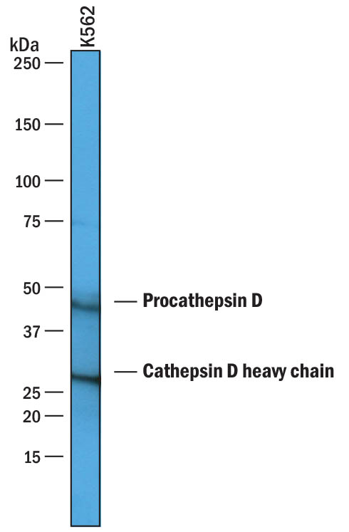
Detection of Human Cathepsin D by Western Blot. Western blot shows lysates of K562 human chronic myelogenous leukemia cell line. PVDF membrane was probed with 1 µg/mL of Goat Anti-Human Cathepsin D Antigen Affinity-purified Polyclonal Antibody (Catalog # AF1014) followed by HRP-conjugated Anti-Goat IgG Secondary Antibody (Catalog # HAF019). Specific bands were detected for Procathepsin D at approximately 45 kDa and Cathepsin D heavy chain 28 kDa (as indicated). This experiment was conducted under reducing conditions and using Immunoblot Buffer Group 1.
 View Larger
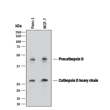
Detection of Human Cathepsin D by Western Blot. Western blot shows lysates of PANC-1 human pancreatic carcinoma cell line and MCF-7 human breast cancer cell line. PVDF membrane was probed with 1 µg/mL of Goat Anti-Human Cathepsin D Antigen Affinity-purified Polyclonal Antibody (Catalog # AF1014) followed by HRP-conjugated Anti-Goat IgG Secondary Antibody (Catalog # HAF017). Specific bands were detected for Procathepsin D at approximately 45 kDa and Cathepsin D heavy chain 28 kDa (as indicated). This experiment was conducted under reducing conditions and using Immunoblot Buffer Group 1.
 View Larger
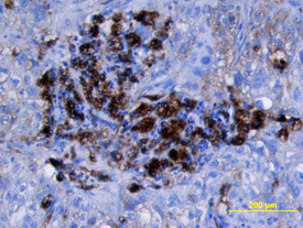
Cathepsin D in Human Lung Cancer Tissue. Cathepsin D was detected in immersion fixed paraffin-embedded sections of human lung cancer tissue using Goat Anti-Human Cathepsin D Antigen Affinity-purified Polyclonal Antibody (Catalog # AF1014) at 15 µg/mL overnight at 4 °C. Tissue was stained using the Anti-Goat HRP-DAB Cell & Tissue Staining Kit (brown; Catalog # CTS008) and counterstained with hematoxylin (blue). View our protocol for Chromogenic IHC Staining of Paraffin-embedded Tissue Sections.
 View Larger
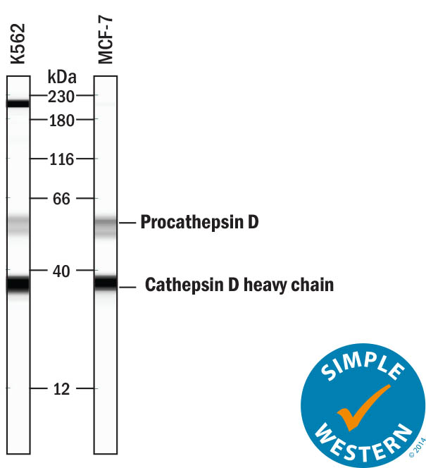
Detection of Human Cathepsin D by Simple WesternTM. Simple Western lane view shows lysates of K562 human chronic myelogenous leukemia cell line and MCF-7 human breast cancer cell line, loaded at 0.2 mg/mL. Specific bands were detected for Procathepsin D at approximately 55 kDa and Cathepsin D heavy chain at approximately 37 kDa (as indicated) using 50 µg/mL of Goat Anti-Human Cathepsin D Antigen Affinity-purified Polyclonal Antibody (Catalog # AF1014) followed by 1:50 dilution of HRP-conjugated Anti-Goat IgG Secondary Antibody (Catalog # HAF109). This experiment was conducted under reducing conditions and using the 12-230 kDa separation system. Non-specific interaction with the 230 kDa Simple Western standard may be seen with this antibody.
Reconstitution Calculator
Preparation and Storage
- 12 months from date of receipt, -20 to -70 °C as supplied.
- 1 month, 2 to 8 °C under sterile conditions after reconstitution.
- 6 months, -20 to -70 °C under sterile conditions after reconstitution.
Background: Cathepsin D
Cathepsin D is a lysosomal aspartic protease of the pepsin family (1). Human cathepsin D is synthesized as a precursor protein, consisting of a signal peptide (residues 1‑18), a propeptide (residues 19-64), and a mature chain (residues 65‑412) (2‑4). The mature chain can be processed further to the light (residues 65‑161) and heavy (residues 169‑412) chains. It is expressed in most cells and overexpressed in breast cancer cells (5). It is a major enzyme in protein degradation in lysosomes, and also involved in the presentation of antigenic peptides. Mice deficient in this enzyme showed a progressive atrophy of the intestinal mucosa, a massive destruction of lymphoid organs, and a profound neuronal ceroid lipofucinosis, indicating that cathepsin D is essential for proteolysis of proteins regulating cell growth and tissue homeostasis (6). Cathepsin D secreted from human prostate carcinoma cells are responsible for the generation of angiostatin, a potent endogeneous inhibitor of angiogenesis (6).
- Conner et al. in Handbook of Proteolytic Enzymes Barrett (1998) Academic Press, San Diego, p. 828.
- Faust, et al. (1985) Proc. Natl. Acad. Sci. USA 82:4910.
- Westley and May (1987) Nucl. Acid Res. 15:3773.
- Redecker, et al. (1991) DNA Cell Biol. 10:423.
- Rochefort, et al. (2000) Clin. Chim. Acta. 291:157.
- Tsukuba, et al. (2000) Mol. Cells 10:601.
Product Datasheets
Citations for Human Cathepsin D Antibody
R&D Systems personnel manually curate a database that contains references using R&D Systems products. The data collected includes not only links to publications in PubMed, but also provides information about sample types, species, and experimental conditions.
13
Citations: Showing 1 - 10
Filter your results:
Filter by:
-
Impaired Lysosome Reformation in Chloroquine-Treated Retinal Pigment Epithelial Cells
Authors: Cardoso, MH;Hall, MJ;Burgoyne, T;Fale, P;Storm, T;Escrevente, C;Antas, P;Seabra, MC;Futter, CE;
Investigative ophthalmology & visual science
Species: Human
Sample Types: Whole Cells
Applications: Immunocytochemistry -
Wild-type and pathogenic forms of ubiquilin 2 differentially modulate components of the autophagy-lysosome pathways
Authors: Idera, A;Sharkey, LM;Kurauchi, Y;Kadoyama, K;Paulson, HL;Katsuki, H;Seki, T;
Journal of pharmacological sciences
Species: Human
Sample Types: Cell Lysates
Applications: Western Blot -
An infection-induced oxidation site regulates legumain processing and tumor growth
Authors: Y Kovalyova, DW Bak, EM Gordon, C Fung, JHB Shuman, TL Cover, MR Amieva, E Weerapana, SK Hatzios
Nature Chemical Biology, 2022-03-24;0(0):.
Species: Gerbil
Sample Types: Cell Lysates
Applications: Western Blot -
Palmitic and Stearic Acids Inhibit Chaperone-Mediated Autophagy (CMA) in POMC-like Neurons In Vitro
Authors: R Espinosa, K Gutiérrez, J Rios, F Ormeño, L Yantén, P Galaz-Davi, CA Ramírez-Sa, V Parra, A Albornoz, IE Alfaro, PV Burgos, E Morselli, A Criollo, M Budini
Cells, 2022-03-08;11(6):.
Species: Human
Sample Types: Cell Culture Supernates
Applications: Western Blot -
Individually cultured bovine embryos produce extracellular vesicles that have the potential to be used as non-invasive embryo quality markers
Authors: K Dissanayak, M Nõmm, F Lättekivi, Y Ressaissi, K Godakumara, A Lavrits, G Midekessa, J Viil, R Bæk, MM Jørgensen, S Bhattachar, A Andronowsk, A Salumets, Ü Jaakma, A Fazeli
Theriogenology, 2020-04-04;149(0):104-116.
Species: Mouse
Sample Types:
Applications: Microarray -
Premature termination codon readthrough upregulates progranulin expression and improves lysosomal function in preclinical models of GRN deficiency
Authors: J Frew, A Baradaran-, AD Balgi, X Wu, TD Yan, S Arns, FS Shidmoossa, J Tan, JB Jaquith, KR Jansen-Wes, FC Lynn, FB Gao, L Petrucelli, HH Feldman, IR Mackenzie, M Roberge, HB Nygaard
Mol Neurodegener, 2020-03-16;15(1):21.
Species: Human
Sample Types: Whole Cells
Applications: ICC -
Identification of PIKfyve kinase as a target in multiple myeloma
Authors: C Bonolo De, YX Zhu, N Sepetov, S Romanov, LA Bruins, CX Shi, CK Stein, JL Petit, AN Polito, ME Sharik, EW Meermeier, GJ Ahmann, ID Lopez Arme, J Kruse, PL Bergsagel, M Chesi, N Meurice, E Braggio, AK Stewart
Haematologica, 2019-10-03;0(0):.
Species: Human
Sample Types: Cell Lysates
Applications: Western Blot -
Visualizing the cellular route of entry of a cystine-knot peptide with Xfect transfection reagent by electron microscopy
Authors: X Gao, A De Mazière, DB Iaea, CP Arthur, J Klumperman, C Ciferri, RN Hannoush
Sci Rep, 2019-05-06;9(1):6907.
Species: Human
Sample Types: Whole Cells
Applications: Electron Microscopy -
HSP90 inhibition targets autophagy and induces a CASP9-dependent resistance mechanism in NSCLC
Authors: J Han, LA Goldstein, W Hou, S Chatterjee, TF Burns, H Rabinowich
Autophagy, 2018-03-21;0(0):1-14.
Species: Human
Sample Types: Tissue Homogenates
Applications: Western Blot -
Amphiphilic star PEG-Camptothecin conjugates for intracellular targeting
J Control Release, 2016-09-24;0(0):.
Species: Human
Sample Types: Whole Cells
Applications: IHC-Fr -
The Apaf-1-binding protein Aven is cleaved by Cathepsin D to unleash its anti-apoptotic potential.
Cell Death Differ., 2012-03-02;19(9):1435-45.
Species: Human
Sample Types: Cell Lysates
Applications: Western Blot -
IFN-gamma regulation of vacuolar pH, cathepsin D processing and autophagy in mammary epithelial cells.
Authors: Khalkhali-Ellis Z, Abbott DE, Bailey CM, Goossens W, Margaryan NV, Gluck SL, Reuveni M, Hendrix MJ
J. Cell. Biochem., 2008-09-01;105(1):208-18.
Species: Human
Sample Types: Cell Lysates, Whole Cells
Applications: ICC, Western Blot -
Clathrin is a key regulator of basolateral polarity.
Authors: Deborde S, Perret E, Gravotta D, Deora A, Salvarezza S, Schreiner R, Rodriguez-Boulan E
Nature, 2008-04-10;452(7188):719-23.
Species: Canine
Sample Types: Cell Lysates
Applications: Immunoprecipitation
FAQs
No product specific FAQs exist for this product, however you may
View all Antibody FAQsReviews for Human Cathepsin D Antibody
There are currently no reviews for this product. Be the first to review Human Cathepsin D Antibody and earn rewards!
Have you used Human Cathepsin D Antibody?
Submit a review and receive an Amazon gift card.
$25/€18/£15/$25CAN/¥75 Yuan/¥2500 Yen for a review with an image
$10/€7/£6/$10 CAD/¥70 Yuan/¥1110 Yen for a review without an image


