Human CD1a Antibody Summary
Asp19-Val300 (predicted)
Accession # P06126
Applications
Please Note: Optimal dilutions should be determined by each laboratory for each application. General Protocols are available in the Technical Information section on our website.
Scientific Data
 View Larger
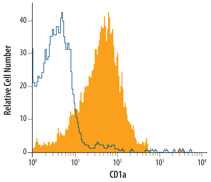
Detection of CD1a in MOLT‑4 Human Cell Line by Flow Cytometry. MOLT-4 human acute lymphoblastic leukemia cell line was stained with Mouse Anti-Human CD1a Monoclonal Antibody (Catalog # MAB7076, filled histogram) or isotype control antibody (MAB002, open histogram), followed by Phycoerythrin-conjugated Anti-Mouse IgG Secondary Antibody (F0102B).
 View Larger
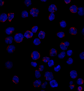
CD1a in MOLT‑4 Human Cell Line. CD1a was detected in immersion fixed MOLT-4 human acute lymphoblastic leukemia cell line using Mouse Anti-Human CD1a Monoclonal Antibody (Catalog # MAB7076) at 10 µg/mL for 3 hours at room temperature. Cells were stained using the NorthernLights™ 557-conjugated Anti-Mouse IgG Secondary Antibody (red; NL007) and counterstained with DAPI (blue). Specific staining was localized to surface and cytoplasm. View our protocol for Fluorescent ICC Staining of Non-adherent Cells.
 View Larger
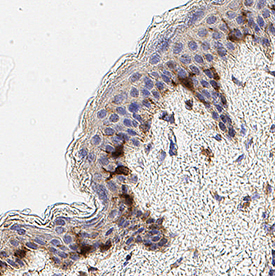
Detection of CD1a in Human Skin. CD1a was detected in immersion fixed paraffin-embedded sections of Human Skin using Mouse Anti-Human CD1a Monoclonal Antibody (Catalog # MAB7076) at 3 µg/mL for 1 hour at room temperature followed by incubation with the Anti-Mouse IgG VisUCyte™ HRP Polymer Antibody (Catalog # VC001). Before incubation with the primary antibody, tissue was subjected to heat-induced epitope retrieval using VisUCyte Antigen Retrieval Reagent-Basic (Catalog # VCTS021). Tissue was stained using DAB (brown) and counterstained with hematoxylin (blue). Specific staining was localized to Langerhan's cells. View our protocol for IHC Staining with VisUCyte HRP Polymer Detection Reagents.
Reconstitution Calculator
Preparation and Storage
- 12 months from date of receipt, -20 to -70 °C as supplied.
- 1 month, 2 to 8 °C under sterile conditions after reconstitution.
- 6 months, -20 to -70 °C under sterile conditions after reconstitution.
Background: CD1a
CD1a is a 49 kDa transmembrane glycoprotein in the CD1 family of glycolipid antigen-presenting MHC-like molecules. CD1a contains one Ig-like domain in its extracellular region. It is expressed by most nonhuman mammals but not by mice or rats. Complexes of CD1a with beta 2-microglobulin and endogenous glycolipids are constitutively expressed on antigen presenting cells, cortical thymocytes, and Langerhans cells. CD1a is a target of autoreactive Th22 helper T cells in the skin.
Product Datasheets
FAQs
No product specific FAQs exist for this product, however you may
View all Antibody FAQsReviews for Human CD1a Antibody
There are currently no reviews for this product. Be the first to review Human CD1a Antibody and earn rewards!
Have you used Human CD1a Antibody?
Submit a review and receive an Amazon gift card.
$25/€18/£15/$25CAN/¥75 Yuan/¥2500 Yen for a review with an image
$10/€7/£6/$10 CAD/¥70 Yuan/¥1110 Yen for a review without an image


