Human CD27/TNFRSF7 Antibody Summary
Thr21-Ile192
Accession # P26842
Applications
Please Note: Optimal dilutions should be determined by each laboratory for each application. General Protocols are available in the Technical Information section on our website.
Scientific Data
 View Larger
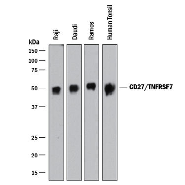
Detection of Human CD27/TNFRSF7 by Western Blot. Western blot shows lysates of Raji human Burkitt's lymphoma cell line, Daudi human Burkitt's lymphoma cell line, Ramos human Burkitt's lymphoma cell line, and human tonsil tissue. PVDF membrane was probed with 1 µg/mL of Goat Anti-Human CD27/TNFRSF7 Antigen Affinity-purified Polyclonal Antibody (Catalog # AF382) followed by HRP-conjugated Anti-Goat IgG Secondary Antibody (HAF017). A specific band was detected for CD27/ TNFRSF7 at approximately 50 kDa (as indicated). This experiment was conducted under reducing conditions and using Immunoblot Buffer Group 1.
 View Larger
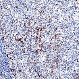
CD27/TNFRSF7 in Human Tonsil. CD27/TNFRSF7 was detected in immersion fixed paraffin-embedded sections of human tonsil using Goat Anti-Human CD27/TNFRSF7 Antigen Affinity-purified Polyclonal Antibody (Catalog # AF382) at 3 µg/mL overnight at 4 °C. Tissue was stained using the Anti-Goat HRP-DAB Cell & Tissue Staining Kit (brown; CTS008) and counterstained with hematoxylin (blue). Specific staining was localized to lymphocytes in germinal center. View our protocol for Chromogenic IHC Staining of Paraffin-embedded Tissue Sections.
 View Larger
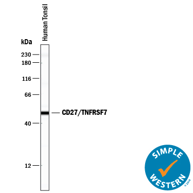
Detection of Human CD27/TNFRSF7 by Simple WesternTM. Simple Western lane view shows lysates of human tonsil tissue, loaded at 0.2 mg/mL. A specific band was detected for CD27/TNFRSF7 at approximately 49 kDa (as indicated) using 20 µg/mL of Goat Anti-Human CD27/ TNFRSF7 Antigen Affinity-purified Polyclonal Antibody (Catalog # AF382) followed by 1:50 dilution of HRP-conjugated Anti-Goat IgG Secondary Antibody (Catalog # HAF109). This experiment was conducted under reducing conditions and using the 12-230 kDa separation system.
 View Larger
CD27/TNFRSF7 Inhibition of Cell Proliferation and Neutralization by Human CD27/TNFRSF7 Antibody. In the presence of sub-optimal amounts of Mouse CD3e Monoclonal Anti-body (Catalog # MAB484) and Recombinant Mouse CD27 Ligand (10 µg/mL, Catalog # 783-CL), Recom-binant Human CD27 Fc Chimera (Catalog # 382-CD) inhibits proliferation in mouse splenic T cells in a dose-dependent manner (orange line). Under these conditions, inhibition of proliferation elicited by Recombinant Human CD27 Fc Chimera (3 µg/mL) is neutralized (green line) by increasing concentrations of Goat Anti-Human CD27 Antigen Affinity-purified Polyclonal Antibody (Catalog # AF382). The ND50 is typically 2.25-9.0 µg/mL.
Reconstitution Calculator
Preparation and Storage
- 12 months from date of receipt, -20 to -70 °C as supplied.
- 1 month, 2 to 8 °C under sterile conditions after reconstitution.
- 6 months, -20 to -70 °C under sterile conditions after reconstitution.
Background: CD27/TNFRSF7
Human CD27 is a lymphocyte-specific member of the TNF receptor superfamily. CD27 is expressed on a subset of human thymocytes and on the majority of mature T cells. CD27 expression is up-regulated after TCR stimulation. Within the CD4+ compartment, it is preferentially expressed on CD45RA+ cells. In contrast, it is preferentially expressed on CD45RO+ cells in the CD8+ compartment. CD27 also appeaars to be a potential marker for memory B cells. It exists as both a disulfide-linked dimer on the cell surface and as a soluble protein found in serum. Human CD27 is a 260 amino acid (aa) protein with a 20 aa signal, a 173 aa extracellular domain, a 20 aa transmembrane domain, and a 47 aa cytoplasmic domain. The ligand for CD27 is CD70. CD70 is expressed on thymic stromal cells and a small subset of activated T cells. Additionally a subset of activated B cells express CD70. The CD27/CD70 interaction appears to be a weak costimulatory pathway involved in T cell and B cell immune response. CD27/CD70 interactions may be more involved in controlling the expansion phase of an immune response. This would be in contrast to B7/CD28 interactions, which are important for the activation phase of immune responses.
- Camerini, D. et al. (1991) J. Immunol. 147:3165.
- Loenen, W.A. et al. (1992) J. Immunol. 149:3937.
- Lens, S.M.A. et al. (1998) Sem. Immunol. 10:491.
Product Datasheets
Citations for Human CD27/TNFRSF7 Antibody
R&D Systems personnel manually curate a database that contains references using R&D Systems products. The data collected includes not only links to publications in PubMed, but also provides information about sample types, species, and experimental conditions.
3
Citations: Showing 1 - 3
Filter your results:
Filter by:
-
Soluble CD27 is an intrathecal biomarker of T-cell-mediated lesion activity in multiple sclerosis
Authors: Cencioni, MT;Magliozzi, R;Palmisano, I;Suwan, K;Mensi, A;Fuentes-Font, L;Villar, LM;Fernández-Velasco, JI;Migallón, NV;Costa-Frossard, L;Monreal, E;Ali, R;Romozzi, M;Mazarakis, N;Reynolds, R;Nicholas, R;Muraro, PA;
Journal of neuroinflammation
Species: Human
Sample Types: Cell Lysates, Whole Tissue
Applications: Immunohistochemistry, Western Blot -
Blockade of protease-activated receptors on T cells correlates with altered proteolysis of CD27 by gingipains of Porphyromonas gingivalis.
Authors: Yun LW, Decarlo AA, Hunter N
Clin. Exp. Immunol., 2007-11-01;150(2):217-29.
Species: Human
Sample Types: Cell Culture Supernates
Applications: ELISA Development -
IL-7 decreases IL-7 receptor alpha (CD127) expression and induces the shedding of CD127 by human CD8+ T cells.
Authors: Vranjkovic A, Crawley AM, Gee K, Kumar A, Angel JB
Int. Immunol., 2007-10-22;19(12):1329-39.
Species: Human
Sample Types: Cell Culture Supernates
Applications: Western Blot
FAQs
No product specific FAQs exist for this product, however you may
View all Antibody FAQsReviews for Human CD27/TNFRSF7 Antibody
Average Rating: 5 (Based on 1 Review)
Have you used Human CD27/TNFRSF7 Antibody?
Submit a review and receive an Amazon gift card.
$25/€18/£15/$25CAN/¥75 Yuan/¥2500 Yen for a review with an image
$10/€7/£6/$10 CAD/¥70 Yuan/¥1110 Yen for a review without an image
Filter by:


