Human CD30/TNFRSF8 Antibody Summary
Phe19-Lys379
Accession # P28908
Applications
This antibody functions as an ELISA detection antibody when paired with Mouse Anti-Human CD30/TNFRSF8 Monoclonal Antibody (Catalog # MAB2291).
This product is intended for assay development on various assay platforms requiring antibody pairs. We recommend the Human CD30/TNFRSF8 DuoSet ELISA Kit (Catalog # DY6126-05) for convenient development of a sandwich ELISA.
The ED50 for this effect is typically 0.2 - 0.6 μg/mL.
Please Note: Optimal dilutions should be determined by each laboratory for each application. General Protocols are available in the Technical Information section on our website.
Scientific Data
 View Larger
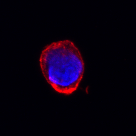
CD30/TNFRSF8 in Jurkat Human Cell Line. CD30/TNFRSF8 was detected in immersion fixed Jurkat human acute T cell leukemia cell line using Goat Anti-Human CD30/TNFRSF8 Antigen Affinity-purified Polyclonal Antibody (Catalog # AF229) at 10 µg/mL for 3 hours at room temperature. Cells were stained using the NorthernLights™ 557-conjugated Anti-Goat IgG Secondary Antibody (red; NL001) and counterstained with DAPI (blue). Specific staining was localized to cell membrane and cytoplasm. View our protocol for Fluorescent ICC Staining of Non-adherent Cells.
 View Larger
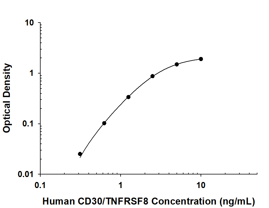
Human CD30/TNFRSF8 ELISA Standard Curve. Recombinant Human CD30/TNFRSF8 protein was serially diluted 2-fold and captured by Mouse Anti-Human CD30/TNFRSF8 Monoclonal Antibody (MAB2291) coated on a Clear Polystyrene Microplate (DY990). Goat Anti-Human CD30/TNFRSF8 Antigen Affinity-purified Polyclonal Antibody (Catalog # AF229) was biotinylated and incubated with the protein captured on the plate. Detection of the standard curve was achieved by incubating Streptavidin-HRP (DY998) followed by Substrate Solution (DY999) and stopping the enzymatic reaction with Stop Solution (DY994).
Reconstitution Calculator
Preparation and Storage
- 12 months from date of receipt, -20 to -70 °C as supplied.
- 1 month, 2 to 8 °C under sterile conditions after reconstitution.
- 6 months, -20 to -70 °C under sterile conditions after reconstitution.
Background: CD30/TNFRSF8
CD30, also known as Ki-1 antigen and TNFRSF8, is a 120 kDa type I transmembrane glycoprotein belonging to the TNF receptor superfamily (1, 2). Mature human CD30 consists of a 361 amino acid (aa) extracellular domain (ECD) with six cysteine-rich repeats, a 28 aa transmembrane segment, and a 188 aa cytoplasmic domain (3). In contrast, mouse and rat CD30 lack 90 aa of the ECD and contain only three cysteine-rich repeats. Within common regions of the ECD, human CD30 shares 53% and 49% aa sequence identity with mouse and rat CD30, respectively. Alternate splicing of human CD30 generates an isoform that includes only the C‑terminal 132 aa of the cytoplasmic domain. CD30 is normally expressed on antigen-stimulated Th cells and B cells (4 - 6). However, it is upregulated in Hodgkin’s disease (on Reed-Sternberg cells), other lymphomas, chronic inflammation, and autoimmunity (7). CD30 binds to CD30 Ligand/TNFSF8 which is expressed on activated Th cells, monocytes, granulocytes and medullary thymic epithelial cells (1, 5). CD30 signaling costimulates antigen-induced Th0 and Th2 proliferation and cytokine secretion but favors a Th2-biased immune response (8). In the absence of antigenic stimulation, it can still induce T cell expression of IL-13 (9). CD30 contributes to thymic negative selection by inducing the apoptotic cell death of CD4+CD8+ T cells (10, 11). In B cells, CD30 ligation promotes cellular proliferation and antibody production in addition to the expression of CXCR4, CCL3, and CCL5 (5, 12). An 85-90 kDa soluble form of CD30 is shed from the cell surface by TACE-mediated cleavage (13, 14). Soluble CD30 retains the ability to bind CD30 Ligand and functions as an inhibitor of normal CD30 signaling (15).
- Kennedy, M.K. et al. (2006) Immunology 118:143.
- Tarkowski, M. (2003) Curr. Opin. Hematol. 10:267.
- Durkop, H. et al. (1992) Cell 68:421.
- Hamann, D. et al. (1996) J. Immunol. 156:1387.
- Shanebeck, S.D. et al. (1995) Eur. J. Immunol. 25:2147.
- Gruss, H.-J. et al. (1994) Blood 83:2045.
- Oflzoglu E. et al. (2009) Adv. Exp. Med. Biol. 647:174.
- Del Prete, G. et al. (1995) J. Exp. Med. 182:1655.
- Harlin, H. et al. (2002) J. Immunol. 169:2451.
- Amakawa, R. et al. (1996) Cell 84:551.
- Chiarle, R. et al. (1999) J. Immunol. 163:194.
- Vinante, F. et al. (2002) Blood 99:52.
- Hansen, H.P. et al. (1995) Int. J. Cancer 63:750.
- Hansen, H.P. et al. (2000) J. Immunol. 165:6703.
- Hargreaves, P.G. and A. Al-Shamkhani (2002) Eur. J. Immunol. 32:163.
Product Datasheets
FAQs
No product specific FAQs exist for this product, however you may
View all Antibody FAQsReviews for Human CD30/TNFRSF8 Antibody
There are currently no reviews for this product. Be the first to review Human CD30/TNFRSF8 Antibody and earn rewards!
Have you used Human CD30/TNFRSF8 Antibody?
Submit a review and receive an Amazon gift card.
$25/€18/£15/$25CAN/¥75 Yuan/¥2500 Yen for a review with an image
$10/€7/£6/$10 CAD/¥70 Yuan/¥1110 Yen for a review without an image


