Human CD81 Antibody Summary
Phe113-Ala201
Accession # P60033
Applications
Please Note: Optimal dilutions should be determined by each laboratory for each application. General Protocols are available in the Technical Information section on our website.
Scientific Data
 View Larger
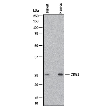
Detection of Human CD81 by Western Blot. Western blot shows lysates of Jurkat human acute T cell leukemia cell line and Ramos human Burkitt's lymphoma cell line. PVDF membrane was probed with 4 µg/mL of Rabbit Anti-Human CD81 Monoclonal Antibody (Catalog # MAB46152) followed by HRP-conjugated Anti-Rabbit IgG Secondary Antibody (HAF008). A specific band was detected for CD81 at approximately 26 kDa (as indicated). This experiment was conducted under reducing conditions and using Western Blot Buffer Group 1.
 View Larger
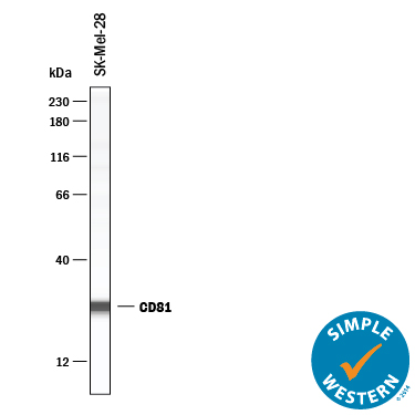
Detection of Human CD81 by Simple WesternTM. Simple Western lane view shows lysates of SK‑Mel‑28 human malignant melanoma cell line, loaded at 0.2 mg/mL. A specific band was detected for CD81 at approximately 27 kDa (as indicated) using 10 µg/mL of Rabbit Anti-Human CD81 Monoclonal Antibody (Catalog # MAB46152). This experiment was conducted under reducing conditions and using the 12-230 kDa separation system.
Reconstitution Calculator
Preparation and Storage
- 12 months from date of receipt, -20 to -70 °C as supplied.
- 1 month, 2 to 8 °C under sterile conditions after reconstitution.
- 6 months, -20 to -70 °C under sterile conditions after reconstitution.
Background: CD81
CD81, also known as TAPA-1 and Tetraspanin-28, is an approximately 25 kDa palmitoylated component of plasma membrane lipid rafts (1). It contains four transmembrane segments, two extracellular loops of 30 and 90 amino acids (aa), and three short cytoplasmic regions (2, 3). Within the large extracellular loop, human CD81 shares 90% and 84% aa sequence identity with mouse and rat CD81, respectively. CD81 associates with a wide range of membrane proteins including CD151, TfR2, LDL R, PCSK9, Glypican 3, IFITM1, IGSF8/CD316, FPRP, and complexes of CD19-CD21 (4-11). It is required for the development of CD4+CD8+ DP thymocytes (12) and hepatocyte infection by Plasmodium sporozoites (13). It also supports B cell receptor signaling (11), Hepcidin expression (5), monocyte and B cell tethering to the vascular endothelium (14), and the immunosuppressive function of Treg and MDSC (15). CD81 additionally functions as a receptor for the E2 glycoprotein of hepatitis C virus (16). The CD81-E2 interaction inhibits NK cell cytolytic activity, provides a co-stimulatory signal to T cells, and inhibits the maturation of plasmacytoid dendritic cells (17-19).
- Charrin, S. et al. (2014) J. Cell Sci. 127:3641.
- Oren, R. et al. (1990) Mol. Cell. Biol. 10:4007.
- Levy, S. et al. (1991) J. Biol. Chem. 266:14597.
- Zhu, Y.-Z. et al. (2012) Virology 429:112.
- Chen, J. and C.A. Enns (2015) J. Biol. Chem. 290:7841.
- Le, Q.-T. et al. (2015) J. Biol. Chem. 290:23385.
- Takahashi, S. et al. (1990) J. Immunol. 145:2207.
- Stipp, C.S. et al. (2001) J. Biol. Chem. 276:40545.
- Stipp, C.S. et al. (2001) J. Biol. Chem. 276:4853.
- Liu, B. et al. (2009) Am. J. Pathol. 175:717.
- Cherukuri, A. et al. (2004) J. Immunol. 279:31973.
- Boismenu, R. et al. (1996) Science 271:198.
- Silvie, O. et al. (2003) Nat. Med. 9:93.
- Feigelson, S.W. et al. (2003) J. Biol. Chem. 278:51203.
- Vences-Catalan, F. et al. (2015) Cancer Res. 75:4517.
- Pileri, P. et al. (1998) Science 282:938.
- Crotta, S. et al. (2002) J. Exp. Med. 195:35.
- Tseng, C.-T. and G.R. Klimpel (2002) J. Exp. Med. 195:43.
- Tu, Z. et al. (2013) Cell. Immunol. 284:98.
Product Datasheets
FAQs
No product specific FAQs exist for this product, however you may
View all Antibody FAQsReviews for Human CD81 Antibody
There are currently no reviews for this product. Be the first to review Human CD81 Antibody and earn rewards!
Have you used Human CD81 Antibody?
Submit a review and receive an Amazon gift card.
$25/€18/£15/$25CAN/¥75 Yuan/¥2500 Yen for a review with an image
$10/€7/£6/$10 CAD/¥70 Yuan/¥1110 Yen for a review without an image

