Human CEACAM-1/CD66a PE-conjugated Antibody Summary
Gln35-Gly428
Accession # P13688
Applications
Please Note: Optimal dilutions should be determined by each laboratory for each application. General Protocols are available in the Technical Information section on our website.
Scientific Data
 View Larger
Detection of CEACAM‑1/CD66a in Human Blood Granulocytes by Flow Cytometry. Human peripheral blood granulocytes were stained with Mouse Anti-Human Fc gamma RIII (CD16) CFS-conjugated Monoclonal Antibody (Catalog # FAB2546F) and either (A) Mouse Anti-Human CEACAM-1/CD66a PE-conjugated Monoclonal Antibody (Catalog # FAB2244P) or (B) Mouse IgG1Phycoerythrin Isotype Control (Catalog # IC002P). View our protocol for Staining Membrane-associated Proteins.
Reconstitution Calculator
Preparation and Storage
- 12 months from date of receipt, 2 to 8 °C as supplied.
Background: CEACAM-1/CD66a
Carcinoembryonic antigen (CEA)-related cell adhesion molecule 1 (CEACAM-1; also BGP) is a 160 kDa member of the CEACAM branch of the CEA gene family of the immunoglobulin superfamily (1-3). It is one of seven human CEACAM subfamily genes that are essentially divided equally between type I transmembrane proteins (CEACAM-1, 3, and 4) and GPI-linked molecules (CEACAM-5-8). There is no CEACAM-2 in human. The gene for human CEACAM-1 codes for a 526 amino acid (aa) type I transmembrane protein that contains a 34 aa signal sequence, a 394 aa extracellular domain (ECD), a 24 aa transmembrane segment, and a 74 aa cytoplasmic region (4, 5). The ECD contains one N-terminal V-type Ig-like domain, followed by three C2-type Ig-like domains. It shows considerable glycosylation, including high mannose residues and (sialyl) LewisX (1). The cytoplasmic region shows one ITIM motif and a calmodulin binding site (1-3). In addition to the full length form, ten alternate splice forms have been reported (1, 4, 6-8). There are three soluble and seven transmembrane isoforms, with variations occurring in both the ECD and cytoplasmic region. All ten alternate splice forms contain the V-type Ig-like domain (aa’s 35-142). The three soluble forms also contain the first two C2-type Ig-like domains (aa’s 145-317), with differences coming in the third C2-type Ig-like domain (6). The seven transmembrane isoforms are highly divergent. Five of the seven contain the V-type plus the first two C2-type domains and then diverge considerably both in the ECD and cytoplasmic region. The remaining two contain only the V‑type Ig-like domain, the transmembrane region, and either a full-length or truncated cytoplasmic tail (1, 8). The actual functions of the isoforms are unclear. Full-length mouse and rat CEACAM-1 are approximately 57% aa identical to human CEACAM-1; in the V‑type Ig-like domain, they are 58% and 56% aa identical, respectively. The full-length molecule is found on neutrophils, bile duct epithelium, activated NK cells, colonic columnar epithelium and endothelium. It is known to act as an intercellular adhesion molecule, forming both homotypic, and heterotypic bonds with CEA and CEACAM-6/NCA (3, 9). On neutrophils, CEACAM-1 also binds to dendritic cell CD-SIGN via its LeX moiety, inducing dendritic cell maturation and a subsequent Th1-type response (10,11).
- Beauchemin, N. et al. (1999) Exp. Cell Res. 252:243.
- Thompson, J. et al. (1992) Genomics 12:761.
- Waggener, C. and S. Ergun (2000) Exp. Cell Res. 261:19.
- Barnett, T.R. et al. (1989) J. Cell Biol. 108:267.
- Hinoda, Y. et al. (1988) Proc. Natl. Acad. Sci. USA 85:6959.
- Kuroki, M. et al. (1991) Biochem. Biophys. Res. Commun. 176:578.
- Barnett, T.R. et al. (1993) Mol. Cell. Biol. 13:1273.
- Watt, S.M. et al. (1994) Blood 84:200.
- Oikawa, S. et al. (1992) Biochem. Biophys. Res. Commun. 186:881.
- Klaas, P.J.M. et al. (2005) FEBS Lett. 579:6159.
- Bogoevska, V. et al. (2005) Glycobiology 16:197.
Product Datasheets
Citations for Human CEACAM-1/CD66a PE-conjugated Antibody
R&D Systems personnel manually curate a database that contains references using R&D Systems products. The data collected includes not only links to publications in PubMed, but also provides information about sample types, species, and experimental conditions.
10
Citations: Showing 1 - 10
Filter your results:
Filter by:
-
Integrated proteomics and transcriptomics analyses identify novel cell surface markers of HIV latency
Authors: Beliakova-Bethell N, Manousopoulou A, Deshmukh S et al.
Virology
-
CEACAM1 is a novel culture-compatible surface marker of expanded long-term reconstituting hematopoietic stem cells
Authors: Unain Ansari, Elisa Tomellini, Jalila Chagraoui, Bernhard Lehnertz, Nadine Mayotte, Marie-Eve Bordeleau et al.
Blood Advances
-
Proinflammatory and Cancer-Promoting Pathobiont Fusobacterium nucleatum Directly Targets Colorectal Cancer Stem Cells
Authors: V Cavallucci, I Palucci, M Fidaleo, A Mercuri, L Masi, V Emoli, G Bianchetti, ME Fiori, G Bachrach, F Scaldaferr, G Maulucci, G Delogu, G Pani
Biomolecules, 2022-09-07;12(9):.
Species: Human
Sample Types: Whole Cells
Applications: Flow Cytometry -
Interferon regulatory factor 1 and a variant of heterogeneous nuclear ribonucleoprotein L coordinately silence the gene for adhesion protein CEACAM1
Authors: KJ Dery, C Silver, L Yang, JE Shively
J. Biol. Chem., 2018-05-02;0(0):.
Species: Human
Sample Types: Whole Cells
Applications: Flow Cytometry -
Assessing the Impact of Targeting CEACAM1 in Head and Neck Squamous Cell Carcinoma
Authors: K Tam, DW Schoppy, JH Shin, JK Tay, U Moreno-Nie, DC Mundy, JB Sunwoo
Otolaryngol Head Neck Surg, 2018-02-13;0(0):1945998187566.
Species: Human
Sample Types: Whole Cells
Applications: Flow Cytometry -
Establishment and Characterization of a Pair of Patient-derived Human Non-small Cell Lung Cancer Cell Lines from a Primary Tumor and Corresponding Lymph Node Metastasis
Authors: F Gebauer, D Wicklein, M Tachezy, T Grob, D Steinemann, G Manukjan, G Göhring, B Schlegelbe, H Maar, JR Izbicki, M Bockhorn, U Schumacher
Anticancer Res, 2016-04-01;36(4):1507-18.
Species: Human
Sample Types: Whole Cells
Applications: Flow Cytometry -
Moraxella catarrhalis adhesin UspA1-derived recombinant fragment rD-7 induces monocyte differentiation to CD14+CD206+ phenotype.
Authors: Xie, Qi, Brackenbury, Louise S, Hill, Darryl J, Williams, Neil A, Qu, Xun, Virji, Mumtaz
PLoS ONE, 2014-03-05;9(3):e90999.
Species: Human
Sample Types: Whole Cells
Applications: Flow Cytometry -
Phenotyping of human melanoma cells reveals a unique composition of receptor targets and a subpopulation co-expressing ErbB4, EPO-R and NGF-R.
Authors: Mirkina I, Hadzijusufovic E, Krepler C, Mikula M, Mechtcheriakova D, Strommer S, Stella A, Jensen-Jarolim E, Holler C, Wacheck V, Pehamberger H, Valent P
PLoS ONE, 2014-01-29;9(1):e84417.
Species: Human
Sample Types: Whole Cells
Applications: Flow Cytometry -
CEACAM1 regulates Fas-mediated apoptosis in Jurkat T-cells via its interaction with beta-catenin.
Authors: Li Y, Shively J
Exp Cell Res, 2013-03-07;319(8):1061-72.
Species: Human
Sample Types: Whole Cells
Applications: Flow Cytometry -
CEACAM 1, 3, 5 and 6 -positive classical monocytes correlate with interstitial lung disease in early systemic sclerosis
Authors: Kana Yokoyama, Hiroki Mitoma, Shotaro Kawano, Yusuke Yamauchi, Qiaolei Wang, Masahiro Ayano et al.
Frontiers in Immunology
FAQs
No product specific FAQs exist for this product, however you may
View all Antibody FAQsReviews for Human CEACAM-1/CD66a PE-conjugated Antibody
Average Rating: 5 (Based on 1 Review)
Have you used Human CEACAM-1/CD66a PE-conjugated Antibody?
Submit a review and receive an Amazon gift card.
$25/€18/£15/$25CAN/¥75 Yuan/¥1250 Yen for a review with an image
$10/€7/£6/$10 CAD/¥70 Yuan/¥1110 Yen for a review without an image
Filter by:
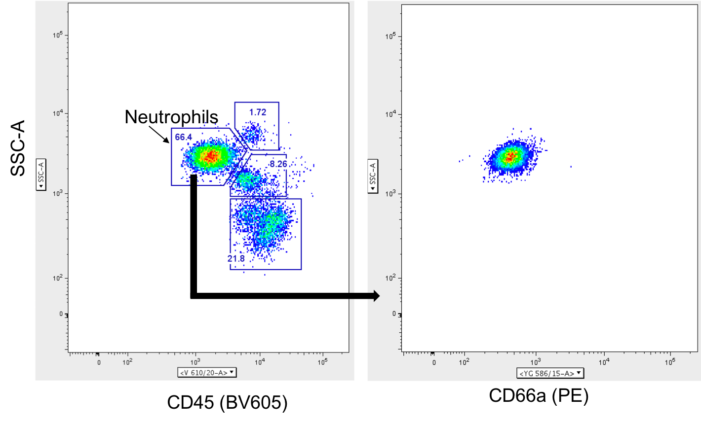
This antibody was used to stain human peripheral whole blood for analysis by flow cytometry. Neutrophils were identified from total live CD45+ leukocytes and expression of CD66a was measured in the PE channel. Consistent fluorescence is observed after fixation in 4% PFA. Good antibody for staining granulocytes


