Human CIS-1 Antibody Summary
Leu11-Leu258
Accession # Q9NSE2
Applications
Please Note: Optimal dilutions should be determined by each laboratory for each application. General Protocols are available in the Technical Information section on our website.
Scientific Data
 View Larger
Detection of Human CIS-1 by Western Blot. Western blot shows lysates of Nalm-6 human Pre-B acute lymphocytic leukemia cell line. PVDF membrane was probed with 1 µg/mL of Goat Anti-Human CIS-1 Antigen Affinity-purified Polyclonal Antibody (Catalog # AF3194) followed by HRP-conjugated Anti-Goat IgG Secondary Antibody (Catalog # HAF019). A specific band was detected for CIS-1 at approximately 35-40 kDa (as indicated). This experiment was conducted under reducing conditions and using Immunoblot Buffer Group 8.
 View Larger
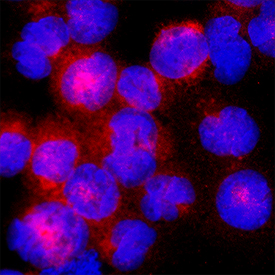
CIS‑1 in HDLM‑2 Human Cell Line. CIS‑1 was detected in immersion fixed HDLM‑2 human Hodgkin’s lymphoma cell line using Goat Anti-Human CIS‑1 Antigen Affinity-purified Polyclonal Antibody (Catalog # AF3194) at 15 µg/mL for 3 hours at room temperature. Cells were stained using the NorthernLights™ 557-conjugated Anti-Goat IgG Secondary Antibody (red; NL001) and counterstained with DAPI (blue). Specific staining was localized to cytoplasm. Staining was performed using our protocol for Fluorescent ICC Staining of Non-adherent Cells.
 View Larger
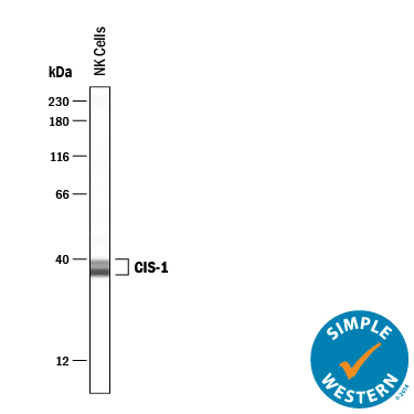
Detection of Human CIS‑1 by Simple WesternTM. Simple Western lane view shows lysates of NK human natural killer lymphoma cell line, loaded at 0.2 mg/mL. Specific bands were detected for CIS‑1 at approximately 37 and 42 kDa (as indicated) using 10 µg/mL of Goat Anti-Human CIS‑1 Antigen Affinity-purified Polyclonal Antibody (Catalog # AF3194). This experiment was conducted under reducing conditions and using the 12-230 kDa separation system.
Reconstitution Calculator
Preparation and Storage
- 12 months from date of receipt, -20 to -70 °C as supplied.
- 1 month, 2 to 8 °C under sterile conditions after reconstitution.
- 6 months, -20 to -70 °C under sterile conditions after reconstitution.
Background: CIS-1
Cytokine Inducible SH2-containing protein (CIS-1) is a 29 kDa protein found in a variety of cell types. Mono or polyubiquitination generally results in a 37 or 45 kDa molecule. CIS-1 binds to phosphorylated cytokine receptors IL-3 R beta and EPO-R and blocks downstream activation of STAT5 via receptor internalization and ubiquitin‑mediated proteosomal degradation. Human CIS-1 is a 258 aa peptide that contains one SH2 domain (aa 82‑163) and one SOCS box (aa 218‑258). There are two known alternatively spliced variants with a 7- or 13-aa substitution for the 7 N-terminal amino acid residues. Over the region used as immunogen, human CIS-1 is 91% identical to the corresponding mouse and canine protein sequences.
Product Datasheets
FAQs
No product specific FAQs exist for this product, however you may
View all Antibody FAQsReviews for Human CIS-1 Antibody
There are currently no reviews for this product. Be the first to review Human CIS-1 Antibody and earn rewards!
Have you used Human CIS-1 Antibody?
Submit a review and receive an Amazon gift card.
$25/€18/£15/$25CAN/¥75 Yuan/¥2500 Yen for a review with an image
$10/€7/£6/$10 CAD/¥70 Yuan/¥1110 Yen for a review without an image

