Human Desmoglein-2 Antibody Summary
Ala50-Gly608 (predicted)
Accession # CAA81226
Applications
Please Note: Optimal dilutions should be determined by each laboratory for each application. General Protocols are available in the Technical Information section on our website.
Scientific Data
 View Larger
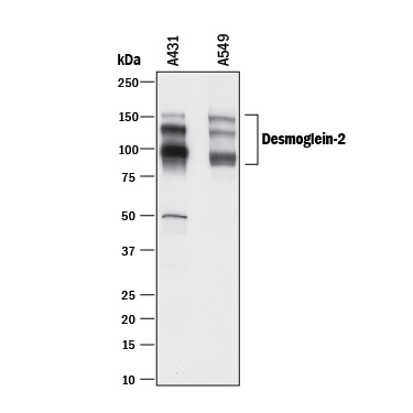
Detection of Human Desmoglein‑2 by Western Blot. Western blot shows lysates of A431 human epithelial carcinoma cell line and A549 human lung carcinoma cell line. PVDF membrane was probed with 0.5 µg/mL of Mouse Anti-Human Desmoglein-2 Monoclonal Antibody (Catalog # MAB947) followed by HRP-conjugated Anti-Mouse IgG Secondary Antibody (Catalog # HAF018). Specific bands were detected for Desmoglein-2 at approximately 90-160 kDa (as indicated). This experiment was conducted under reducing conditions and using Immunoblot Buffer Group 1.
 View Larger
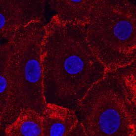
Desmoglein‑2 in NHEK Human Cells. Desmoglein-2 was detected in immersion fixed NHEK human normal epidermal keratinocytes using Mouse Anti-Human Desmoglein-2 Monoclonal Antibody (Catalog # MAB947) at 10 µg/mL for 3 hours at room temperature. Cells were stained using the NorthernLights™ 557-conjugated Anti-Mouse IgG Secondary Antibody (red; Catalog # NL007) and counterstained with DAPI (blue). Specific staining was localized to cytoplasm and cell junctions. View our protocol for Fluorescent ICC Staining of Cells on Coverslips.
 View Larger
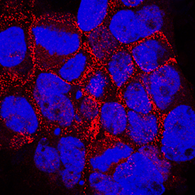
Desmoglein‑2 in A431 Human Cell Line. Desmoglein-2 was detected in immersion fixed A431 human epithelial carcinoma cell line wildtype and knockout using Mouse Anti-Human Desmoglein-2 Monoclonal Antibody (Catalog # MAB947) at 10 µg/mL for 3 hours at room temperature. Cells were stained using the NorthernLights™ 557-conjugated Anti-Mouse IgG Secondary Antibody (red; Catalog # NL007) and counterstained with DAPI (blue). Specific staining was localized to plasma membrane. View our protocol for Fluorescent ICC Staining of Cells on Coverslips.
 View Larger
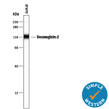
Detection of Human Desmoglein‑2 by Simple WesternTM. Simple Western lane view shows lysates of Jurkat human acute T cell leukemia cell line, loaded at 0.2 mg/mL. A specific band was detected for Desmoglein‑2 at approximately 120 kDa (as indicated) using 20 µg/mL of Mouse Anti-Human Desmoglein‑2 Monoclonal Antibody (Catalog # MAB947). This experiment was conducted under reducing conditions and using the 12-230 kDa separation system.
Reconstitution Calculator
Preparation and Storage
- 12 months from date of receipt, -20 to -70 °C as supplied.
- 1 month, 2 to 8 °C under sterile conditions after reconstitution.
- 6 months, -20 to -70 °C under sterile conditions after reconstitution.
Background: Desmoglein-2
Desmoglein-2 is one of three members of the desmoglein subfamily of calcium-dependent cadherin cell adhesion molecules. Together with desmocollins, another subfamily within the cadherin superfamily, the desmoglein isoforms form the adhesive components of desmosomes, the cell-cell adhesive structures that are found in epithelial cells. Human Desmoglein-2 is a type I transmembrane glycoprotein of 1117 amino acid (aa) residues with a 23 aa signal peptide and a 25 aa propeptide. It differs from other classic cadherins by having four instead of five cadherin repeat domains in its extracellular region, and a much larger cytoplasmic region containing five desmoglein repeat domains which share homology with the cadherin repeats. Instead of having the HAV adhesion motif found in type I cadherins, desmogleins have R/YAL as the adhesion motif on its amino-terminal cadherin repeat. The cytoplasmic tails of desmogleins interact with desmoplakins, plakoglobin and plakophilins. In turn, these proteins link the desmogleins with the intermediate filaments. Desmoglein-2 has been shown to be important in establishing cell-cell adhesion and function in epithelial cells. Desmoglein-2 was originally identified in colon carcinoma and colon, and was named HDGC (human desmoglein colon).
- Nollet, R. et al. (2000) J. Mol. Biol. 299:551.
- Elias, P. et al. (2001) J. Cell Biol. 153:243.
- Arnemann, J. et al. (1992) Genomics 13:484.
Product Datasheets
Citation for Human Desmoglein-2 Antibody
R&D Systems personnel manually curate a database that contains references using R&D Systems products. The data collected includes not only links to publications in PubMed, but also provides information about sample types, species, and experimental conditions.
1 Citation: Showing 1 - 1
-
Prospective isolation and global gene expression analysis of definitive and visceral endoderm.
Authors: Sherwood RI, Jitianu C, Cleaver O, Shaywitz DA, Lamenzo JO, Chen AE, Golub TR, Melton DA
Dev. Biol., 2007-01-12;304(2):541-55.
Species: Mouse
Sample Types: Whole Tissue
Applications: IHC
FAQs
No product specific FAQs exist for this product, however you may
View all Antibody FAQsReviews for Human Desmoglein-2 Antibody
Average Rating: 5 (Based on 2 Reviews)
Have you used Human Desmoglein-2 Antibody?
Submit a review and receive an Amazon gift card.
$25/€18/£15/$25CAN/¥75 Yuan/¥2500 Yen for a review with an image
$10/€7/£6/$10 CAD/¥70 Yuan/¥1110 Yen for a review without an image
Filter by:


