Human Endophilin B1/B2 Antibody Summary
Ala33-Asn189
Accession # Q9Y371
Applications
Please Note: Optimal dilutions should be determined by each laboratory for each application. General Protocols are available in the Technical Information section on our website.
Scientific Data
 View Larger
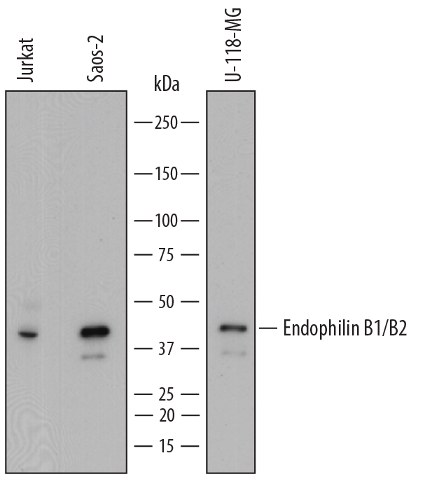
Detection of Human Endophilin B1/B2 by Western Blot. Western blot shows lysates of Jurkat human acute T cell leukemia cell line, Saos-2 human osteosarcoma cell line, and U-118-MG human glioblastoma/astrocytoma cell line. PVDF membrane was probed with 0.2 µg/mL of Mouse Anti-Human Endophilin B1/B2 Monoclonal Antibody (Catalog # MAB7456) followed by HRP-conjugated Anti-Mouse IgG Secondary Antibody (Catalog # HAF018). A specific band was detected for Endophilin B1/B2 at approximately 43 kDa (as indicated). This experiment was conducted under reducing conditions and using Immunoblot Buffer Group 1.
 View Larger
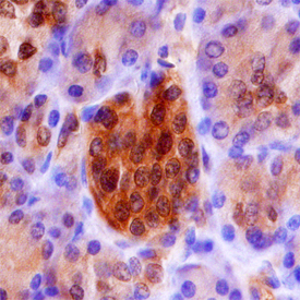
Endophilin B1/B2 in Human Pancreas. Endophilin B1/B2 was detected in immersion fixed paraffin-embedded sections of human pancreas using Mouse Anti-Human Endophilin B1/B2 Monoclonal Antibody (Catalog # MAB7456) at 15 µg/mL overnight at 4 °C. Before incubation with the primary antibody, tissue was subjected to heat-induced epitope retrieval using Antigen Retrieval Reagent-Basic (Catalog # CTS013). Tissue was stained using the Anti-Mouse HRP-DAB Cell & Tissue Staining Kit (brown; Catalog # CTS002) and counter-stained with hematoxylin (blue). Specific staining was localized to cytoplasm of islet cells. View our protocol for Chromogenic IHC Staining of Paraffin-embedded Tissue Sections.
 View Larger
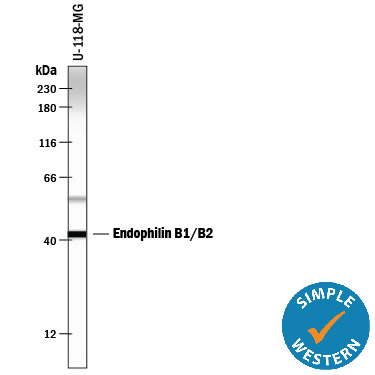
Detection of Human Endophilin B1/B2 by Simple WesternTM. Simple Western lane view shows lysates of U‑118‑MG human glioblastoma/astrocytoma cell line, loaded at 0.5 mg/mL. A specific band was detected for Endophilin B1/B2 at approximately 42 kDa (as indicated) using 2 µg/mL of Mouse Anti-Human Endophilin B1/B2 Monoclonal Antibody (Catalog # MAB7456). This experiment was conducted under reducing conditions and using the 12-230 kDa separation system.
 View Larger
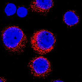
Endophilin B1/B2 in Jurkat Human Cell Line. Endophilin B1/B2 was detected in immersion fixed Jurkat human acute T cell leukemia cell line using Mouse Anti-Human Endophilin B1/B2 Monoclonal Antibody (Catalog # MAB7456) at 8 µg/mL for 3 hours at room temperature. Cells were stained using the NorthernLights™ 557-conjugated Anti-Mouse IgG Secondary Antibody (red; Catalog # NL007) and counterstained with DAPI (blue). Specific staining was localized to cytoplasm. View our protocol for Fluorescent ICC Staining of Non-adherent Cells.
Reconstitution Calculator
Preparation and Storage
- 12 months from date of receipt, -20 to -70 °C as supplied.
- 1 month, 2 to 8 °C under sterile conditions after reconstitution.
- 6 months, -20 to -70 °C under sterile conditions after reconstitution.
Background: Endophilin B1/B2
Endophilins are among the best known BAR (Bin/Amphiphysin/Rvs) domain proteins. The highly conserved protein dimerisation domain is typically involved in membrane dynamics. Endophilin-B1, also known as SH3GLB1 (Src-Homology 3 Domain-containing GRB2-like protein B1) and BIF-1, is a 40 kDa protein that belongs to the endophilin family of molecules. It is a cytoplasmic and Golgi membrane protein that is expressed in neurons, striated (skeletal and cardiac) muscle cells, and placenta. Using SH3GLB1 in yeast two-hybrid screens, a second protein, SH3GLB2 (endophilin B2), was identified as an interacting partner. Endophilin-B1 plays a role in cell homeostasis. In neurons, Endophilin-B1 is phosphorylated by Cdk5, inducing homodimerization and autophagosome formation. In addition, it appears to play a role in the maintenance of mitochondrial integrity. Endophilin-B1 appears to cycle on-and-off the outer mitochondrial membrane (OMM), contributing to OMM integrity. And after the initiation of endocytosis, it also directs EEA1+ TrkA-containing endosomes back into the cell membrane for reuse. Human Endophilin-B1 is 365 amino acids (aa) in length. It possesses an N-terminal lipid-binding BAR (Bin/Amphiphysin/Rvs) domain (aa 27-261) that contains an internal coiled-coil region, and a C‑terminal SH3 domain that binds Pro-rich sequences on potential ligands (309-363). There are at least three potential splice variants for Endophilin-B1. Two are 42 and 43 kDa in size, and possess a 21 and 29 aa insertion after Ser190, respectively. A third potentially utilizes an alternative start site at Met101. Over aa 33-189, human Endophilin-B1 shares 98% aa sequence identity with mouse Endophilin-B1.
Product Datasheets
FAQs
No product specific FAQs exist for this product, however you may
View all Antibody FAQsReviews for Human Endophilin B1/B2 Antibody
Average Rating: 5 (Based on 1 Review)
Have you used Human Endophilin B1/B2 Antibody?
Submit a review and receive an Amazon gift card.
$25/€18/£15/$25CAN/¥75 Yuan/¥2500 Yen for a review with an image
$10/€7/£6/$10 CAD/¥70 Yuan/¥1110 Yen for a review without an image
Filter by:


