Human EPCR Antibody Summary
Ser18-Ser210
Accession # Q9UNN8
Applications
Please Note: Optimal dilutions should be determined by each laboratory for each application. General Protocols are available in the Technical Information section on our website.
Scientific Data
 View Larger
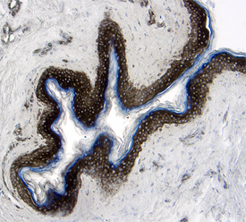
EPCR in Human Skin. EPCR was detected in immersion fixed paraffin-embedded sections of human skin using 25 µg/mL Mouse Anti-Human EPCR Monoclonal Antibody (Catalog # MAB22451) overnight at 4 °C. Tissue was stained with the Anti-Mouse HRP-DAB Cell & Tissue Staining Kit (brown; Catalog # CTS002) and counterstained with hematoxylin (blue). View our protocol for Chromogenic IHC Staining of Paraffin-embedded Tissue Sections.
Reconstitution Calculator
Preparation and Storage
- 12 months from date of receipt, -20 to -70 °C as supplied.
- 1 month, 2 to 8 °C under sterile conditions after reconstitution.
- 6 months, -20 to -70 °C under sterile conditions after reconstitution.
Background: EPCR
Protein C is a vitamin K-dependent serine protease that plays a major role in blood coagulation. Binding of Protein C to EPCR (endothelial protein C receptor) leads to the proteolytic activation of PAR1 (protease-activated receptor 1) on endothelial cells and subsequent up-regulation of Protein C-induced genes. EPCR is a type I transmembrane glycoprotein in the CD1/MHC family. It is expressed most strongly in the endothelial cells of arteries and veins in heart and lung. Membrane bound EPCR is released by metalloproteolytic cleavage to generate the soluble receptor. The extracellular domain of human and mouse EPCR shares approximately 61% amino acid sequence homology.
Product Datasheets
Citations for Human EPCR Antibody
R&D Systems personnel manually curate a database that contains references using R&D Systems products. The data collected includes not only links to publications in PubMed, but also provides information about sample types, species, and experimental conditions.
2
Citations: Showing 1 - 2
Filter your results:
Filter by:
-
Identification of two major autoantigens negatively regulating endothelial activation in Takayasu arteritis
Authors: T Mutoh, T Shirai, T Ishii, Y Shirota, F Fujishima, F Takahashi, Y Kakuta, Y Kanazawa, A Masamune, Y Saiki, H Harigae, H Fujii
Nat Commun, 2020-03-09;11(1):1253.
Species: Human
Sample Types: Cell Culture Lysates
Applications: Western Blot -
Endothelial protein C receptor is overexpressed in colorectal cancer as a result of amplification and hypomethylation of chromosome 20q
Authors: N Lal, CR Willcox, A Beggs, P Taniere, A Shikotra, P Bradding, R Adams, D Fisher, G Middleton, C Tselepis, BE Willcox
J Pathol Clin Res, 2017-07-14;3(3):155-170.
Species: Human
Sample Types: Whole Tissue
Applications: IHC-P
FAQs
No product specific FAQs exist for this product, however you may
View all Antibody FAQsReviews for Human EPCR Antibody
There are currently no reviews for this product. Be the first to review Human EPCR Antibody and earn rewards!
Have you used Human EPCR Antibody?
Submit a review and receive an Amazon gift card.
$25/€18/£15/$25CAN/¥75 Yuan/¥2500 Yen for a review with an image
$10/€7/£6/$10 CAD/¥70 Yuan/¥1110 Yen for a review without an image

