Human EPCR Antibody Summary
Ser18-Ser210
Accession # Q9UNN8
Applications
Please Note: Optimal dilutions should be determined by each laboratory for each application. General Protocols are available in the Technical Information section on our website.
Scientific Data
 View Larger
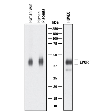
Detection of Human EPCR by Western Blot. Western blot shows lysates of human skin tissue, human placenta tissue, and HUVEC human umbilical vein endothelial cells. PVDF membrane was probed with 0.25 µg/mL of Goat Anti-Human EPCR Antigen Affinity-purified Polyclonal Antibody (Catalog # AF2245) followed by HRP-conjugated Anti-Goat IgG Secondary Antibody (Catalog # HAF017). A specific band was detected for EPCR at approximately 40-45 kDa (as indicated). This experiment was conducted under reducing conditions and using Immunoblot Buffer Group 1.
 View Larger
EPCR in Human Liver. EPCR was detected in immersion fixed paraffin-embedded sections of human liver using 1.7 µg/mL Goat Anti-Human EPCR Antigen Affinity-purified Polyclonal Antibody (Catalog # AF2245) overnight at 4 °C. Tissue was stained with the Anti-Goat HRP-DAB Cell & Tissue Staining Kit (brown; Catalog # CTS008) and counterstained with hematoxylin (blue). View our protocol for Chromogenic IHC Staining of Paraffin-embedded Tissue Sections.
Reconstitution Calculator
Preparation and Storage
- 12 months from date of receipt, -20 to -70 °C as supplied.
- 1 month, 2 to 8 °C under sterile conditions after reconstitution.
- 6 months, -20 to -70 °C under sterile conditions after reconstitution.
Background: EPCR
EPCR is a type I transmembrane glycoprotein in the CD1/MHC family. It is expressed most strongly in the endothelial cells of arteries and veins in heart and lung. Membrane bound EPCR is released by metalloproteolytic cleavage to generate the soluble receptor. Protein C is a vitamin K-dependent serine protease that plays a major role in blood coagulation. Binding of Protein C to EPCR leads to the proteolytic activation of PAR1 (protease-activated receptor 1) on endothelial cells and subsequent up-regulation of Protein C-induced genes. The extracellular domain of human and mouse EPCR shares approximately 61% amino acid sequence homology.
Product Datasheets
Citations for Human EPCR Antibody
R&D Systems personnel manually curate a database that contains references using R&D Systems products. The data collected includes not only links to publications in PubMed, but also provides information about sample types, species, and experimental conditions.
10
Citations: Showing 1 - 10
Filter your results:
Filter by:
-
Non-myogenic mesenchymal cells contribute to muscle degeneration in facioscapulohumeral muscular dystrophy patients
Authors: L Di Pietro, F Giacalone, E Ragozzino, V Saccone, F Tiberio, M De Bardi, M Picozza, G Borsellino, W Lattanzi, E Guadagni, S Bortolani, G Tasca, E Ricci, O Parolini
Cell Death & Disease, 2022-09-16;13(9):793.
Species: Human
Sample Types: Whole Tissue
Applications: IHC -
EPCR-PAR1 biased signaling regulates perfusion recovery and neovascularization in peripheral ischemia
Authors: ML Bochenek, R Gogiraju, S Gro beta mann, J Krug, J Orth, S Reyda, GS Georgiadis, H Spronk, S Konstantin, T Münzel, JH Griffin, PS Wild, C Espinola-K, W Ruf, K Schäfer
JCI Insight, 2022-07-22;0(0):.
Species: Human, Mouse
Sample Types: Cell Lysates, Whole Tissue
Applications: IHC, Western Blot -
Interplay of Plasmodium falciparum and thrombin in brain endothelial barrier disruption
Authors: M Avril, M Benjamin, MM Dols, JD Smith
Sci Rep, 2019-09-11;9(1):13142.
Species: Human
Sample Types: Whole Cells
Applications: Flow Cytometry -
Blood outgrowth endothelial cells (BOECs) as a novel tool for studying adhesion of Plasmodium falciparum-infected erythrocytes
Authors: G Ecklu-Mens, RW Olsen, A Bengtsson, MF Ofori, L Hviid, ATR Jensen, Y Adams
PLoS ONE, 2018-10-09;13(10):e0204177.
Species: Human
Sample Types: Whole Cells
Applications: Flow Cytometry, ICC -
Infected erythrocytes expressing DC13 PfEMP1 differ from recombinant proteins in EPCR-binding function
Authors: Y Azasi, G Lindergard, A Ghumra, J Mu, LH Miller, JA Rowe
Proc. Natl. Acad. Sci. U.S.A., 2018-01-16;0(0):.
Species: Human
Sample Types: Whole Cells
Applications: Binding Assay -
Interaction between Endothelial Protein C Receptor and Intercellular Adhesion Molecule 1 to Mediate Binding of Plasmodium falciparum-Infected Erythrocytes to Endothelial Cells
MBio, 2016-07-12;7(4):.
Species: Human
Sample Types: Whole Cells
Applications: Flow Cytometry -
Cellular regulation of blood coagulation: a model for venous stasis.
Authors: Campbell JE, Brummel-Ziedins KE, Butenas S
Blood, 2010-09-23;116(26):6082-91.
Species: Human
Sample Types: Whole Cells
Applications: ICC -
Comprehensive analysis of conditioned media from ovarian cancer cell lines identifies novel candidate markers of epithelial ovarian cancer.
Authors: Gunawardana CG, Kuk C, Smith CR
J. Proteome Res., 2009-10-01;8(10):4705-13.
Species: Human
Sample Types: Cell Culture Supernates
Applications: ELISA Development -
Aldosterone modifies hemostasis via upregulation of the protein-C receptor in human vascular endothelium.
Authors: Ducros E, Berthaut A, Mirshahi SS, Faussat AM, Soria J, Agarwal MK, Mirshahi M
Biochem. Biophys. Res. Commun., 2008-06-13;373(2):192-6.
Species: Human
Sample Types: Cell Lysates, Whole Cells
Applications: Dot Blot, Flow Cytometry, ICC -
Protein C is an autocrine growth factor for human skin keratinocytes.
Authors: Xue M, Campbell D, Jackson CJ
J. Biol. Chem., 2007-02-09;282(18):13610-6.
Species: Human
Sample Types: Whole Tissue
Applications: IHC-P
FAQs
No product specific FAQs exist for this product, however you may
View all Antibody FAQsReviews for Human EPCR Antibody
There are currently no reviews for this product. Be the first to review Human EPCR Antibody and earn rewards!
Have you used Human EPCR Antibody?
Submit a review and receive an Amazon gift card.
$25/€18/£15/$25CAN/¥75 Yuan/¥2500 Yen for a review with an image
$10/€7/£6/$10 CAD/¥70 Yuan/¥1110 Yen for a review without an image

