Human G-CSF Antibody

Catalog # Availability Size / Price Qty
MAB2141-100
MAB2141-SP
Detection of G‑CSF in U‑87 MG (Positive) & MOLT‑4 (Negative).
2 Images
Product Details
FAQs
Supplemental Products
Reviews

Human G-CSF Antibody Summary

Species Reactivity
Human
Specificity
Detects human G-CSF by direct ELISA.
Source
Monoclonal Mouse IgG1 Clone # 741250
Purification
Protein A or G purified from hybridoma culture supernatant
Immunogen
E. coli-derived human G-CSF protein
Thr31-Leu204
Accession # P09919
Formulation
Lyophilized from a 0.2 μm filtered solution in PBS with Trehalose. *Small pack size (SP) is supplied either lyophilized or as a 0.2 µm filtered solution in PBS.
Label
Unconjugated

Applications

Recommended Concentration
Sample
ELISA
This antibody functions as an ELISA capture antibody whenpaired with human anti G-CSF Monoclonal Antibody (Catalog # MAB11296).Thisproduct is intended for assay development on various assay platforms requiringantibody pairs.
 
Immunocytochemistry
8 µg/mL
Immersion fixed U‑87 MG human glioblastoma/astrocytoma cell line (Positive) & MOLT‑4 human acute lymphoblastic leukemia cell line (Negative)

Please Note: Optimal dilutions should be determined by each laboratory for each application. General Protocols are available in the Technical Information section on our website.

Scientific Data

Immunocytochemistry View Larger

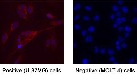
Detection of G‑CSF in U‑87 MG (Positive) & MOLT‑4 (Negative). G‑CSF was detected in immersion fixed U‑87 MG human glioblastoma/astrocytoma cell line (Positive) & absent in MOLT‑4 human acute lymphoblastic leukemia cell line (Negative) using Mouse Anti-Human G‑CSF Monoclonal Antibody (Catalog # MAB2141) at 8 µg/mL for 3 hours at room temperature. Cells were stained using the NorthernLights™ 557-conjugated Anti-Mouse IgG Secondary Antibody (red; Catalog # NL007) and counterstained with DAPI (blue). Specific staining was localized to Cell surface and cytoplasm. View our protocol for Fluorescent ICC Staining of Cells on Coverslips.

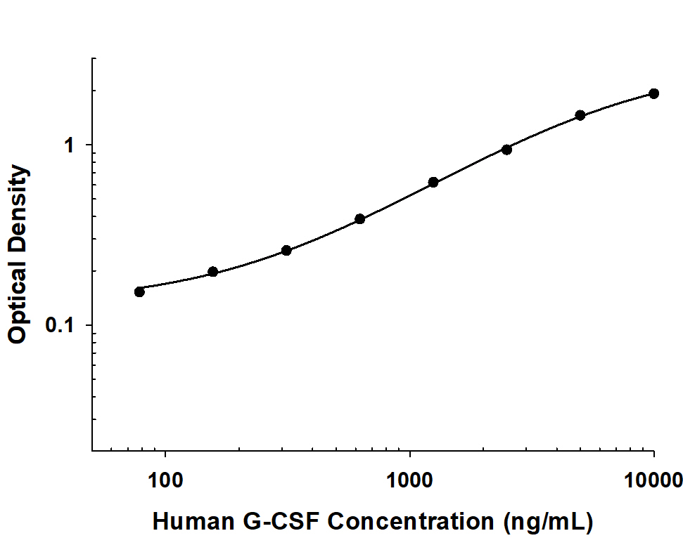
Human G‑CSF ELISA Standard Curve. Recombinant Human G‑CSF protein was serially diluted 2-fold and captured by Mouse Anti-Human G‑CSF Monoclonal Antibody (Catalog # MAB2141) coated on a Clear Polystyrene Microplate (Catalog # DY990). Mouse Anti-Human G‑CSF Monoclonal Antibody (MAB11296) was biotinylated and incubated with the protein captured on the plate. Detection of the standard curve was achieved by incubating Streptavidin-HRP (Catalog # DY998) followed by Substrate Solution (Catalog # DY999) and stopping the enzymatic reaction with Stop Solution (Catalog # DY994).

Reconstitution Calculator

Reconstitution Calculator

The reconstitution calculator allows you to quickly calculate the volume of a reagent to reconstitute your vial. Simply enter the mass of reagent and the target concentration and the calculator will determine the rest.

=
÷

Preparation and Storage

Reconstitution
Reconstitute at 0.5 mg/mL in sterile PBS.
Loading...
Shipping
The product is shipped at ambient temperature. Upon receipt, store it immediately at the temperature recommended below. *Small pack size (SP) is shipped with polar packs. Upon receipt, store it immediately at -20 to -70 °C
Stability & Storage
Use a manual defrost freezer and avoid repeated freeze-thaw cycles.
  • 12 months from date of receipt, -20 to -70 °C as supplied.
  • 1 month, 2 to 8 °C under sterile conditions after reconstitution.
  • 6 months, -20 to -70 °C under sterile conditions after reconstitution.

Background: G-CSF

G-CSF is a pleiotropic cytokine best known for its specific effects on the proliferation, differentiation, and activation of hematopoietic cells of the neutrophilic granulocyte lineage. It is produced mainly by monocytes and macrophages upon activation by endotoxin, TNF-alpha and IFN-gamma. Other cell types including fibroblasts, endothelial cells, astrocytes and bone marrow stromal cells can also secrete G-CSF after LPS, IL-1 or TNF-alpha activation. In addition, various carcinoma cell lines and myeloblastic leukemia cells can express G-CSF constitutively.
In humans, two distinct cDNA clones for G-CSF, encoding 207 and 204 amino acid precursor proteins, have been isolated. Both proteins have a 30 amino acid signal peptide and have identical amino acid sequences except for a three amino acid insertion (deletion) at the 35th amino acid residue from the N-terminus of the mature protein. Human G-CSF is 73% identical at the amino acid level to murine G-CSF and the two proteins show species cross-reactivity.
In vitro, G-CSF stimulates growth, differentiation and functions of cells from the neutrophil lineage. It also has blast cell growth factor activity and can synergize with IL-3 to shorten the Go period of early hematopoietic progenitors. Consistent with its in vitro functions, G-CSF has been found to play important roles in defense against infection, in inflammation and repair, and in the maintenance of steady state hematopoiesis.

Long Name
Granulocyte Colony Stimulating Factor
Entrez Gene IDs
1440 (Human); 12985 (Mouse)
Alternate Names
C17orf33; chromosome 17 open reading frame 33; colony stimulating factor 3 (granulocyte); CSF3; CSF3OS; Filgrastim; GCSF; G-CSF; GCSFlenograstim; granulocyte colony-stimulating factor; Lenograstim; MGC45931; Pluripoietin

Product Datasheets

You must select a language.

x

FAQs

No product specific FAQs exist for this product, however you may

View all Antibody FAQs
Loading...

Reviews for Human G-CSF Antibody

There are currently no reviews for this product. Be the first to review Human G-CSF Antibody and earn rewards!

Have you used Human G-CSF Antibody?

Submit a review and receive an Amazon gift card.

$25/€18/£15/$25CAN/¥75 Yuan/¥1250 Yen for a review with an image

$10/€7/£6/$10 CAD/¥70 Yuan/¥1110 Yen for a review without an image

Submit a Review