Human Glut2 Antibody Summary
Applications
Please Note: Optimal dilutions should be determined by each laboratory for each application. General Protocols are available in the Technical Information section on our website.
Scientific Data
 View Larger
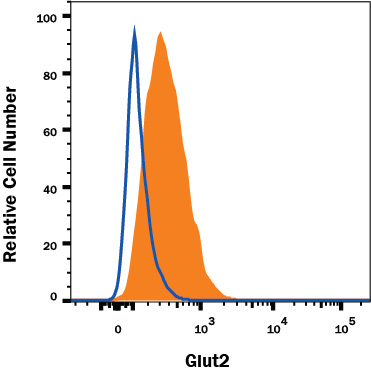
Detection of Glut2 in HepG2 Human Cell Line by Flow Cytometry. HepG2 human hepatocellular carcinoma cell line was stained with Mouse Anti-Human Glut2 Monoclonal Antibody (Catalog # MAB1414, filled histogram) or isotype control antibody (Catalog # MAB003, open histogram), followed by Phycoerythrin-conjugated Anti-Mouse IgG Secondary Antibody (Catalog # F0102B). View our protocol for Staining Membrane-associated Proteins.
 View Larger
Glut2 in Human Pancreatic Cancer Tissue. Glut2 was detected in immersion fixed paraffin-embedded sections of human pancreatic cancer tissue using Mouse Anti-Human Glut2 Monoclonal Antibody (Catalog # MAB1414) at 5 µg/mL overnight at 4 °C. Tissue was stained using the Anti-Mouse HRP-DAB Cell & Tissue Staining Kit (brown; Catalog # CTS002) and counterstained with hematoxylin (blue). Specific labeling was localized to the plasma membrane of exocrine cells. View our protocol for Chromogenic IHC Staining of Paraffin-embedded Tissue Sections.
 View Larger
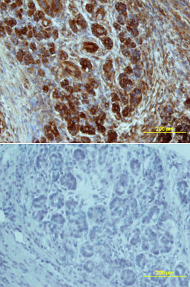
Glut2 in Human Pancreas. Glut2 was detected in immersion fixed paraffin-embedded sections of human pancreas array using Mouse Anti-Human Glut2 Monoclonal Antibody (Catalog # MAB1414) at 25 µg/mL overnight at 4 °C. Tissue was stained using the Anti-Mouse HRP-DAB Cell & Tissue Staining Kit (brown; Catalog # CTS002) and counterstained with hematoxylin (blue). Lower panel shows a lack of labeling if primary antibodies are omitted and tissue is stained only with secondary antibody followed by incubation with detection reagents. View our protocol for Chromogenic IHC Staining of Paraffin-embedded Tissue Sections.
Reconstitution Calculator
Preparation and Storage
- 12 months from date of receipt, -20 to -70 degreesC as supplied. 1 month, 2 to 8 degreesC under sterile conditions after reconstitution. 6 months, -20 to -70 degreesC under sterile conditions after reconstitution.
Background: Glut2
Glut2 belongs to the facilitative glucose transporter protein family that comprises 13 members. It is an integral membrane protein with 12 transmembrane domains. Glut2 is expressed predominantly in liver, intestine, kidney, and pancreatic beta-cells. It is a low-affinity glucose transporter that has been suggested to function as a glucose sensor in pancreatic beta-cells and facilitate either glucose uptake or efflux from cells depending on the nutritional state (1).
- Olson, A.L. and J.E. Pessin (1996) Annu. Rev. Nut. 16:235.
- Mahraoui, L. et al. (1994) J. Biochem. 298:629.
Product Datasheets
Citations for Human Glut2 Antibody
R&D Systems personnel manually curate a database that contains references using R&D Systems products. The data collected includes not only links to publications in PubMed, but also provides information about sample types, species, and experimental conditions.
4
Citations: Showing 1 - 4
Filter your results:
Filter by:
-
Mutations in SLC2A2 gene reveal hGLUT2 function in pancreatic beta cell development.
Authors: Michau A, Guillemain G, Grosfeld A, Vuillaumier-Barrot S, Grand T, Keck M, L'Hoste S, Chateau D, Serradas P, Teulon J, De Lonlay P, Scharfmann R, Brot-Laroche E, Leturque A, Le Gall M
J Biol Chem, 2013-08-28;288(43):31080-92.
Species: Human, Xenopus
Sample Types: Oocyte, Whole Cells
Applications: IHC -
Pancreatic islet cell phenotype and endocrine function throughout diabetes development in non-obese diabetic mice.
Authors: Kornete, Mara, Beauchemin, Hugues, Polychronakos, Constant, Piccirillo, Ciriaco
Autoimmunity, 2013-02-04;46(4):259-68.
Species: Mouse
Sample Types: Whole Cells
Applications: Flow Cytometry -
Ongoing beta-cell turnover in adult nonhuman primates is not adaptively increased in streptozotocin-induced diabetes.
Authors: Saisho Y, Manesso E, Butler AE, Galasso R, Kavanagh K, Flynn M, Zhang L, Clark P, Gurlo T, Toffolo GM, Cobelli C, Wagner JD, Butler PC
Diabetes, 2011-01-26;60(3):848-56.
Species: Primate - Chlorocebus aethiops (African Green Monkey)
Sample Types: Whole Tissue
Applications: IHC-P -
Isolation and characterization of a stem cell population from adult human liver.
Authors: Herrera MB, Bruno S, Buttiglieri S, Tetta C, Gatti S, Deregibus MC, Bussolati B, Camussi G
Stem Cells, 2006-08-31;24(12):2840-50.
Species: Human
Sample Types: Whole Cells
Applications: ICC
FAQs
No product specific FAQs exist for this product, however you may
View all Antibody FAQsReviews for Human Glut2 Antibody
There are currently no reviews for this product. Be the first to review Human Glut2 Antibody and earn rewards!
Have you used Human Glut2 Antibody?
Submit a review and receive an Amazon gift card.
$25/€18/£15/$25CAN/¥75 Yuan/¥2500 Yen for a review with an image
$10/€7/£6/$10 CAD/¥70 Yuan/¥1110 Yen for a review without an image


