Human LAP (TGF-beta 1) Antibody Summary
Leu30-Ser390
Accession # P01137
Applications
Please Note: Optimal dilutions should be determined by each laboratory for each application. General Protocols are available in the Technical Information section on our website.
Scientific Data
 View Larger
Detection of Human LAP (TGF-beta 1) by Western Blot. Western blot shows lysates of Saos-2 human osteosarcoma cell line. PVDF membrane was probed with 2 µg/mL of Goat Anti-Human LAP (TGF-beta 1) Antigen Affinity-purified Polyclonal Antibody (Catalog # AF-246-NA) followed by HRP-conjugated Anti-Goat IgG Secondary Antibody (Catalog # HAF017). A specific band was detected for LAP (TGF-beta 1) at approximately 48 kDa (as indicated). This experiment was conducted under reducing conditions and using Immunoblot Buffer Group 1.
 View Larger
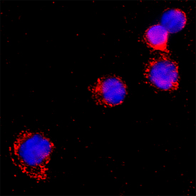
LAP (TGF-beta 1) in Human PBMCs. LAP (TGF-beta 1) was detected in immersion fixed Human peripheral blood mononuclear cells using Goat Anti-Human LAP (TGF-beta 1) Antigen Affinity-purified Polyclonal Antibody (Catalog # AF-246-NA) at 15 µg/mL for 3 hours at room temperature. Cells were stained using the NorthernLights™ 557-conjugated Anti-Goat IgG Secondary Antibody (red; NL001) and counterstained with DAPI (blue). Specific staining was localized to cytoplasm. Staining was performed using our protocol for Fluorescent ICC Staining of Non-adherent Cells.
 View Larger
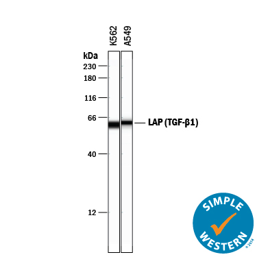
Detection of Human LAP (TGF-beta 1) by Simple WesternTM. Simple Western lane view shows lysates of K562 human chronic myelogenous leukemia cell line and A549 human lung carcinoma cell line, loaded at 0.2 mg/mL. A specific band was detected for LAP (TGF-beta 1) at approximately 61 kDa (as indicated) using 20 µg/mL of Goat Anti-Human LAP (TGF-beta 1) Antigen Affinity-purified Polyclonal Antibody (Catalog # AF-246-NA) followed by 1:50 dilution of HRP-conjugated Anti-Goat IgG Secondary Antibody (Catalog # HAF109). This experiment was conducted under reducing conditions and using the 12-230 kDa separation system.
 View Larger
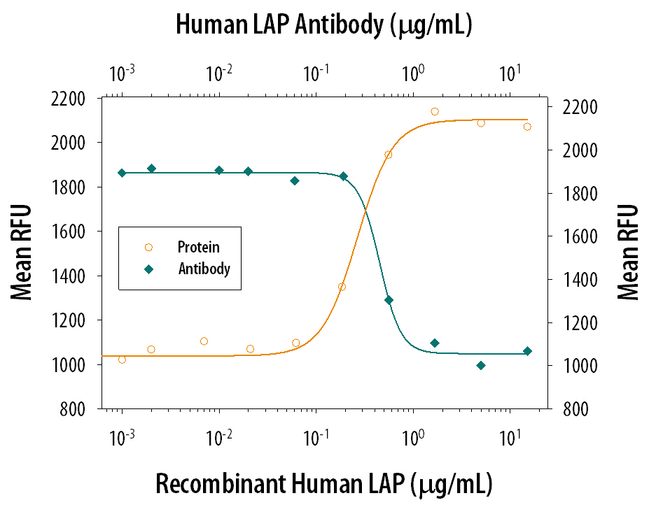
LAP TGF‑ beta 1 Inhibition of TGF‑ beta 1 Activity and Neutralization by Human LAP TGF‑ beta 1 Antibody. Recombinant Human LAP TGF-beta 1 (Catalog # 246-LP) inhibits Recom-binant Human TGF-beta 1 (Catalog # 240-B) growth inhibition activity in the HT-2 mouse T cell line in a dose-dependent manner (orange line). Inhibition of Recom-binant Human TGF-beta 1 (1 ng/mL) activity elicited by Recombin-ant Human LAP TGF-beta 1 (500 ng/mL) is neutralized (green line) by increasing concentrations of Goat Anti-Human LAP TGF-beta 1 Antigen Affinity-purified Polyclonal Antibody (Catalog # AF-246-NA). The ND50 is typically 0.4-2 µg/mL.
Reconstitution Calculator
Preparation and Storage
- 12 months from date of receipt, -20 to -70 °C as supplied.
- 1 month, 2 to 8 °C under sterile conditions after reconstitution.
- 6 months, -20 to -70 °C under sterile conditions after reconstitution.
Background: LAP (TGF-beta 1)
TGF-beta 1 (transforming growth factor beta 1) and the closely related TGF-beta 2 and -beta 3 are members of the large TGF-beta superfamily. TGF‑ beta proteins are highly pleiotropic cytokines that regulate processes such as immune function, proliferation and epithelial-mesenchymal transition (1‑3). Human TGF-beta 1 cDNA encodes a 390 amino acid (aa) precursor that contains a 29 aa signal peptide and a 361 aa proprotein (4). A furin-like convertase processes the proprotein within the trans-Golgi to generate an N‑terminal 249 aa (aa 30-278) latency-associated peptide (LAP) and a C-terminal 112 aa mature TGF-beta 1 (aa 279-390) (4‑6). Disulfide-linked homodimers of LAP and TGF-beta 1 remain non‑covalently associated after secretion, forming the small latent TGF-beta 1 complex (4‑8). Purified LAP is also capable of associating with active TGF‑ beta with high affinity, and can neutralize TGF-beta activity (9). Covalent linkage of LAP to one of three latent TGF-beta binding proteins (LTBPs) creates a large latent complex that may interact with the extracellular matrix (5-7). TGF-beta activation from latency is controlled both spatially and temporally, by multiple pathways that include actions of proteases such as plasmin and MMP9, and/or by thrombospondin 1 or selected integrins (5, 8). The LAP portion of human TGF-beta 1 shares 91%, 92%, 85%, 86% and 88% aa identity with porcine, canine, mouse, rat and equine TGF-beta 1 LAP, respectively, while mature human TGF-beta 1 portion shares 100% aa identity with porcine, canine and bovine TGF-beta 1, and 99% aa identity with mouse, rat and equine TGF-beta 1. Although different isoforms of TGF-beta are naturally associated with their own distinct LAPs, the TGF-beta 1 LAP is capable of complexing with, and inactivating, all other human TGF-beta isoforms and those of most other species (9). Mutations within the LAP are associated with Camurati-Engelmann disease, a rare sclerosing bone dysplasia characterized by inappropriate presence of active TGF‑ beta 1 (10).
- Dunker, N. & K. Krieglstein (2000) Eur. J. Biochem. 267:6982.
- Wahl, S.M. (2006) Immunol. Rev. 213:213.
- Chang, H. et al. (2002) Endocr. Rev. 23:787.
- Derynck, R. et al. (1985) Nature 316:701.
- Dabovic, B. and D.B. Rifkin (2008) “TGF-beta Bioavailability” in The TGF-beta Family. Derynck, R. and K. Miyazono (eds): Cold Spring Harbor Laboratory Press, p. 179.
- Brunner, A.M. et al. (1989) J. Biol. Chem. 264:13660.
- Miyazono, K. et al. (1991) EMBO J. 10:1091.
- Oklu, R. and R. Hesketh (2000) Biochem. J. 352:601.
- Miller, D.M. et al. (1992) Mol. Endocrinol. 6:694.
- Janssens, K. et al. (2003) J. Biol. Chem. 278:7718.
Product Datasheets
Citations for Human LAP (TGF-beta 1) Antibody
R&D Systems personnel manually curate a database that contains references using R&D Systems products. The data collected includes not only links to publications in PubMed, but also provides information about sample types, species, and experimental conditions.
19
Citations: Showing 1 - 10
Filter your results:
Filter by:
-
Heme stimulates platelet mitochondrial oxidant production to induce targeted granule secretion
Authors: GK Annarapu, D Nolfi-Done, M Reynolds, Y Wang, L Kohut, B Zuckerbrau, S Shiva
Redox Biology, 2021-12-05;48(0):102205.
Species: Human
Sample Types: Cell Culture Supernates
Applications: Dot Blot -
Excessive exosome release is the pathogenic pathway linking a lysosomal deficiency to generalized fibrosis
Authors: D van de Vle, J Demmers, XX Nguyen, Y Campos, E Machado, I Annunziata, H Hu, E Gomero, X Qiu, A Bongiovann, CA Feghali-Bo, A d'Azzo
Sci Adv, 2019-07-17;5(7):eaav3270.
Species: Mouse
Sample Types: Exosomes
Applications: Western Blot -
Immune cell expression of TGF?1 in cancer with lymphoid stroma: dendritic cell and regulatory T cell contact
Authors: H Ohtani, T Terashima, E Sato
Virchows Arch., 2018-03-28;0(0):.
Species: Human
Sample Types: Whole Cells
Applications: IHC-P -
Multicolor quantitative confocal imaging cytometry
Authors: DL Coutu, KD Kokkaliari, L Kunz, T Schroeder
Nat. Methods, 2017-11-13;15(1):39-46.
Species: Mouse
Sample Types: Whole Tissue
Applications: IHC -
Cutting Edge: ERK1 Mediates the Autocrine Positive Feedback Loop of TGF-? and Furin in Glioma-Initiating Cells
Authors: E Ventura, M Weller, I Burghardt
J. Immunol., 2017-05-08;0(0):.
Species: Human
Sample Types: Cell Lysates
Applications: Western Blot -
The Use of Platelet-Rich and Platelet-Poor Plasma to Enhance Differentiation of Skeletal Myoblasts: Implications for the Use of Autologous Blood Products for Muscle Regeneration
Authors: O Miroshnych, WT Chang, JL Dragoo
Am J Sports Med, 2016-12-27;45(4):945-953.
Species: Human
Sample Types: Protein
Applications: Western Blot -
MicroRNA-21 preserves the fibrotic mechanical memory of mesenchymal stem cells
Authors: Boris Hinz
Nat Mater, 2016-10-31;0(0):.
Species: Rat
Sample Types: Tissue Homogenates
Applications: Western Blot -
Identification of bone morphogenetic protein 7 (BMP7) as an instructive factor for human epidermal Langerhans cell differentiation.
Authors: Yasmin N, Bauer T, Modak M, Wagner K, Schuster C, Koffel R, Seyerl M, Stockl J, Elbe-Burger A, Graf D, Strobl H
J Exp Med, 2013-11-04;210(12):2597-610.
Species: Human
Sample Types: Whole Tissue
Applications: IHC-Fr -
Secretome and degradome profiling shows that Kallikrein-related peptidases 4, 5, 6, and 7 induce TGFbeta-1 signaling in ovarian cancer cells.
Authors: Shahinian H, Loessner D, Biniossek M, Kizhakkedathu J, Clements J, Magdolen V, Schilling O
Mol Oncol, 2013-10-01;8(1):68-82.
Species: Human, Mouse
Sample Types: Cell Culture Supernates, Whole Tissue
Applications: IHC, Western Blot -
Identification of the thiol isomerase-binding peptide, mastoparan, as a novel inhibitor of shear-induced transforming growth factor beta1 (TGF-beta1) activation.
Authors: Brophy T, Coller B, Ahamed J
J Biol Chem, 2013-03-05;288(15):10628-39.
Species: Human
Sample Types: Cell Lysates
Applications: Western Blot -
TGF-beta mediates homing of bone marrow-derived human mesenchymal stem cells to glioma stem cells.
Authors: Shinojima N, Hossain A, Takezaki T, Fueyo J, Gumin J, Gao F, Nwajei F, Marini F, Andreeff M, Kuratsu J, Lang F
Cancer Res, 2013-01-30;73(7):2333-44.
Species: Human
Sample Types: Whole Cells
Applications: Neutralization -
Nonmyelinating Schwann cells maintain hematopoietic stem cell hibernation in the bone marrow niche.
Authors: Yamazaki S, Ema H, Karlsson G, Yamaguchi T, Miyoshi H, Shioda S, Taketo MM, Karlsson S, Iwama A, Nakauchi H
Cell, 2011-11-23;147(5):1146-58.
Species: Mouse
Sample Types: Whole Cells
Applications: ICC -
Diverse functions of reactive cysteines facilitate unique biosynthetic processes of aggregate-prone interleukin-31.
Authors: Shen M, Siu S, Byrd S, Edelmann KH, Patel N, Ketchem RR, Mehlin C, Arnett HA, Hasegawa H
Exp. Cell Res., 2010-12-21;317(7):976-93.
Species: Human
Sample Types: Cell Lysates
Applications: Western Blot -
Human pregnancy specific beta-1-glycoprotein 1 (PSG1) has a potential role in placental vascular morphogenesis.
Authors: Ha CT, Wu JA, Irmak S, Lisboa FA, Dizon AM, Warren JW, Ergun S, Dveksler GS
Biol. Reprod., 2010-03-24;83(1):27-35.
Species: Human
Sample Types: Whole Cells
Applications: Neutralization -
Transforming growth factor-beta signaling in hypertensive remodeling of porcine aorta.
Authors: Popovic N, Bridenbaugh EA, Neiger JD, Hu JJ, Vannucci M, Mo Q, Trzeciakowski J, Miller MW, Fossum TW, Humphrey JD, Wilson E
Am. J. Physiol. Heart Circ. Physiol., 2009-08-28;297(6):H2044-53.
Species: Porcine
Sample Types: Whole Tissue
Applications: IHC -
Induction of epithelial-mesenchymal transition in primary airway epithelial cells from patients with asthma by transforming growth factor-beta1.
Authors: Hackett TL, Warner SM, Stefanowicz D, Shaheen F, Pechkovsky DV, Murray LA, Argentieri R, Kicic A, Stick SM, Bai TR, Knight DA
Am. J. Respir. Crit. Care Med., 2009-04-30;180(2):122-33.
Species: Human
Sample Types: Whole Cells
Applications: Neutralization -
Transglutaminase 2 limits murine peritoneal acute gout-like inflammation by regulating macrophage clearance of apoptotic neutrophils.
Authors: Rose DM, Sydlaske AD, Agha-Babakhani A, Johnson K, Terkeltaub R
Arthritis Rheum., 2006-10-01;54(10):3363-71.
Species: Mouse
Sample Types: Whole Cells
Applications: Neutralization -
Compromised lymphocytes infiltrate hepatocellular carcinoma: the role of T-regulatory cells.
Authors: Unitt E, Rushbrook SM, Marshall A, Davies S, Gibbs P, Morris LS, Coleman N, Alexander GJ
Hepatology, 2005-04-01;41(4):722-30.
Species: Human
Sample Types: Whole Tissue
Applications: IHC-P -
Cord blood CD4(+)CD25(+)-derived T regulatory cell lines express FoxP3 protein and manifest potent suppressor function.
Authors: Godfrey WR, Spoden DJ, Ge YG, Baker SR, Liu B, Levine BL, June CH, Blazar BR, Porter SB
Blood, 2004-09-16;105(2):750-8.
Species: Human
Sample Types: Cell Culture Supernates
Applications: Neutralization
FAQs
No product specific FAQs exist for this product, however you may
View all Antibody FAQsReviews for Human LAP (TGF-beta 1) Antibody
Average Rating: 5 (Based on 1 Review)
Have you used Human LAP (TGF-beta 1) Antibody?
Submit a review and receive an Amazon gift card.
$25/€18/£15/$25CAN/¥75 Yuan/¥2500 Yen for a review with an image
$10/€7/£6/$10 CAD/¥70 Yuan/¥1110 Yen for a review without an image
Filter by:


