Human MFG-E8 Antibody Summary
Leu24-Cys387
Accession # Q08431
Applications
Please Note: Optimal dilutions should be determined by each laboratory for each application. General Protocols are available in the Technical Information section on our website.
Scientific Data
 View Larger
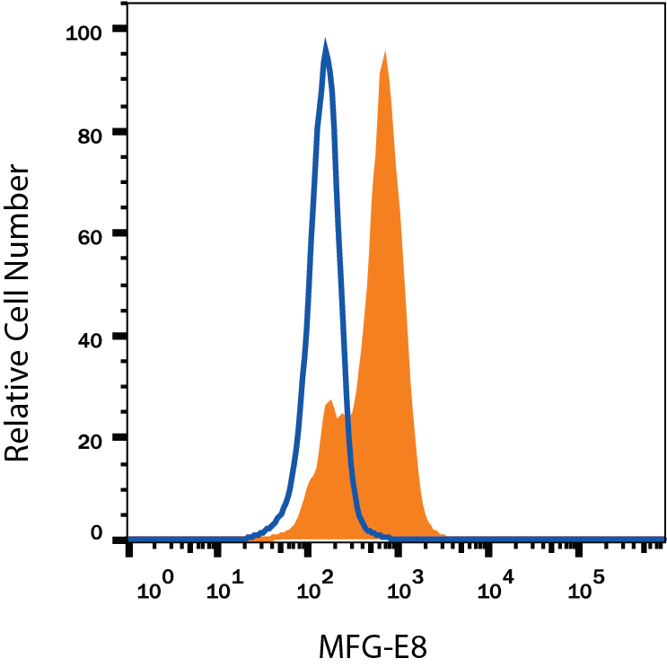
Detection of MFG‑E8 in Human Immature Dendritic Cells by Flow Cytometry. Human immature dendritic cells were stained with Mouse Anti-Human MFG-E8 Monoclonal Antibody (Catalog # MAB27671, filled histogram) or isotype control antibody (MAB003, open histogram), followed by Phycoerythrin-conjugated Anti-Mouse IgG Secondary Antibody (F0102B). To facilitate intracellular staining, cells were fixed with Flow Cytometry Fixation Buffer (FC004) and permeabilized with Flow Cytometry Permeabilization/Wash Buffer I (FC005). View our protocol for Staining Intracellular Molecules.
 View Larger
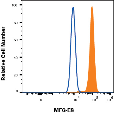
Detection of MFG‑E8 in TF-1 cells by Flow Cytometry. TF-1 cells were stained with Mouse Anti-Human MFG‑E8 Monoclonal Antibody (Catalog # MAB27671, filled histogram) or isotype control antibody (Catalog # MAB003, open histogram), followed by Fluorescein-conjugated Anti-Mouse IgG Secondary Antibody (Catalog # F0103B). To facilitate intracellular staining, cells were fixed with FC012 and permeabilized with FoxP3 Perm. View our protocol for Staining Intracellular Molecules.
Reconstitution Calculator
Preparation and Storage
- 12 months from date of receipt, -20 to -70 °C as supplied.
- 1 month, 2 to 8 °C under sterile conditions after reconstitution.
- 6 months, -20 to -70 °C under sterile conditions after reconstitution.
Background: MFG-E8
Milk Fat Globulin Protein E8 (MFG-E8), also known as Lactadherin, MP47, breast epithelial antigen BA46, and SED1, is a 66‑75 kDa pleiotropic secreted glycoprotein that promotes mammary gland morphogenesis, angiogenesis, and tumor progression. MFG-E8 also plays an important role in tissue homeostasis and the prevention of inflammation (1). Human MGF-E8 contains one N-terminal EGF-like domain and two C‑terminal F5/8-type discoidin-like domains (2). It shares 63% and 61% aa sequence identity with comparable regions of mouse and rat MFG-E8, respectively. Shorter isoforms of human MFG-E8 may have N-terminal deletions (beginning near the end of the first discoidin-like domain), internal deletions (lacking either the EGF-like domain or the central region of the second discoidin-like domain), or C‑terminal deletions (truncated within the second discoidin-like domain) (3). A 50 aa internal proteolytic fragment of human MFG-E8 (known as Medin) is a major component of aortic medial amyloid deposits (4). MFG-E8 is released into the milk in complex with lipid-containing milk fat globules. It is also found in multiple other cell types including endothelial cells and smooth muscle cells of the vasculature, immature dendritic cells, at the acrosomal cap of testicular and epididymal sperm, and in epithelial cells of the endometrium (1). MFG-E8 binds to the Integrins alpha V beta 3 and alpha V beta 5 and potentiates the angiogenic action of VEGF through VEGF R2 (5, 6). It reduces inflammation and tissue damage in a variety of settings. MFG-E8 functions as a bridge between phosphatidylserine on apoptotic cells and Integrin alpha V beta 3 on phagocytes, leading to the clearance of apoptotic debris (7). It mediates the engulfment of apoptotic bodies in atherosclerotic plaques and prion-infected brain (8, 9) and of apoptotic B cells during germinal center reactions (10, 11). MFG-E8 also promotes the removal of excess Collagen in fibrotic lungs and the regeneration of damaged intestinal epithelia (12, 13). Its tissue-protective role impairs anti‑tumor immunity and chemotherapy-induced apoptosis (14). MFG-E8 in the breastmilk blocks rotavirus infection in nursing babies (15).
- Raymond, A. et al. (2009) J. Cell. Biochem. 106:957.
- Couto, J.R. et al. (1996) DNA Cell Biol. 15:281.
- Yamaguchi, H. et al. (2010) Eur. J. Immunol. 40:1778.
- Haggqvist, B. et al. (1999) Proc. Natl. Acad. Sci. USA 96:8669.
- Silvestre, J.-S. et al. (2005) Nat. Med. 11:499.
- Borges, E. et al. (2000) J. Biol. Chem. 275:39867.
- Hanayama, R. et al. (2002) Nature 417:182.
- Ait-Oufella, H. et al. (2007) Circulation 115:2168.
- Kranich, J. et al. (2010) J. Exp. Med. 207:2271.
- Hanayama, R. et al. (2004) Science 304:1147.
- Kranich, J. et al. (2010) J. Exp. Med. 205:1293.
- Atabai, K. et al. (2009) J. Clin. Invest. 119:3713.
- Bu, H.-F. et al. (2007) J. Clin. Invest. 117:3673.
- Jinushi, M. et al. (2009) J. Exp. Med. 206:1317.
- Kvistgaard, A.S. et al. (2004) J. Dairy Sci. 87:4088.
Product Datasheets
Citations for Human MFG-E8 Antibody
R&D Systems personnel manually curate a database that contains references using R&D Systems products. The data collected includes not only links to publications in PubMed, but also provides information about sample types, species, and experimental conditions.
2
Citations: Showing 1 - 2
Filter your results:
Filter by:
-
Ultrafiltration combined with size exclusion chromatography efficiently isolates extracellular vesicles from cell culture media for compositional and functional studies
Authors: BJ Benedikter, FG Bouwman, T Vajen, ACA Heinzmann, G Grauls, EC Mariman, EFM Wouters, PH Savelkoul, C Lopez-Igle, RR Koenen, GGU Rohde, FRM Stassen
Sci Rep, 2017-11-10;7(1):15297.
Species: Human
Sample Types: Cell Lysates
Applications: Western Blot -
Milk fat globule EGF-8 promotes melanoma progression through coordinated Akt and twist signaling in the tumor microenvironment.
Authors: Jinushi M, Nakazaki Y, Carrasco DR, Draganov D, Souders N, Johnson M, Mihm MC, Dranoff G
Cancer Res., 2008-11-01;68(21):8889-98.
Species: Human
Sample Types: Whole Tissue
Applications: IHC-P
FAQs
No product specific FAQs exist for this product, however you may
View all Antibody FAQsReviews for Human MFG-E8 Antibody
Average Rating: 4 (Based on 1 Review)
Have you used Human MFG-E8 Antibody?
Submit a review and receive an Amazon gift card.
$25/€18/£15/$25CAN/¥75 Yuan/¥2500 Yen for a review with an image
$10/€7/£6/$10 CAD/¥70 Yuan/¥1110 Yen for a review without an image
Filter by:


