Human/Mouse Brachyury APC-conjugated Antibody Summary
Ser2-Glu202
Accession # O15178
Applications
Please Note: Optimal dilutions should be determined by each laboratory for each application. General Protocols are available in the Technical Information section on our website.
Scientific Data
 View Larger
Detection of Brachyury in D3 Mouse Cell Line by Flow Cytometry. D3 mouse embryonic stem cell line differentiated with mouse serum for 4 days was stained with Goat Anti-Human/Mouse Brachyury APC-conjugated Antigen Affinity-purified Polyclonal Antibody (Catalog # IC2085A, filled histogram) or isotype control antibody (IC108A, open histogram). To facilitate intracellular staining, cells were fixed with Flow Cytometry Fixation Buffer (FC004) and permeabilized with Flow Cytometry Permeabilization/Wash Buffer I (FC005). View our protocol for Staining Intracellular Molecules.
 View Larger
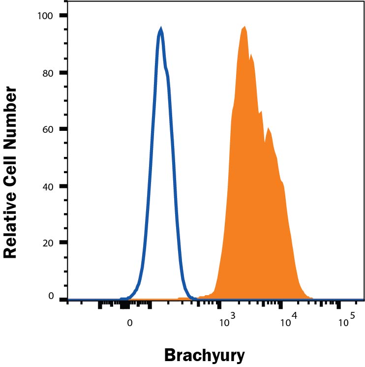
Detection of Brachyury in IPSC mesoderm differentiation cells by Flow Cytometry. IPSC mesoderm differentiation cells treated with IPSC mesoderm differentiation (SC030B) for 2 days were stained with Goat Anti-Human/Mouse Brachyury APC‑conjugated Antigen Affinity-purified Polyclonal Antibody (Catalog # IC2085A, filled histogram) or isotype control antibody (Catalog # IC108A, open histogram). To facilitate intracellular staining, cells were fixed with FC004 and permeabilized with FoxP3 Perm. View our protocol for Staining Intracellular Molecules.
Reconstitution Calculator
Preparation and Storage
- 12 months from date of receipt, 2 to 8 °C as supplied.
Background: Brachyury
Brachyury is the founding member of the T-box family of transcription factors, which is characterized by the N-terminal conserved DNA-binding T-domain. Brachyury is required in the early determination and differentiation of mesoderms. Human brachyury molecule shares 90% homology with mouse brachyury.
Product Datasheets
Citations for Human/Mouse Brachyury APC-conjugated Antibody
R&D Systems personnel manually curate a database that contains references using R&D Systems products. The data collected includes not only links to publications in PubMed, but also provides information about sample types, species, and experimental conditions.
5
Citations: Showing 1 - 5
Filter your results:
Filter by:
-
hPSC-derived sacral neural crest enables rescue in a severe model of Hirschsprung's disease
Authors: Y Fan, J Hackland, A Baggiolini, LY Hung, H Zhao, P Zumbo, P Oberst, AP Minotti, E Hergenrede, S Najjar, Z Huang, NM Cruz, A Zhong, M Sidharta, T Zhou, E de Stanchi, D Betel, RM White, M Gershon, KG Margolis, L Studer
Cell Stem Cell, 2023-03-02;30(3):264-282.e9.
Species: Human, Mouse
Sample Types: Whole Cells
Applications: Flow Cytometry -
hPSC-derived sacral neural crest enables rescue in a severe model of Hirschsprung's disease
Authors: Y Fan, J Hackland, A Baggiolini, LY Hung, H Zhao, P Zumbo, P Oberst, AP Minotti, E Hergenrede, S Najjar, Z Huang, NM Cruz, A Zhong, M Sidharta, T Zhou, E de Stanchi, D Betel, RM White, M Gershon, KG Margolis, L Studer
Cell Stem Cell, 2023;30(3):264-282.e9.
Species: Human
Sample Types: Whole Cells
Applications: Flow Cytometry -
Lateral plate mesoderm cell-based organoid system for NK cell regeneration from human pluripotent stem cells
Authors: D Huang, J Li, F Hu, C Xia, Q Weng, T Wang, H Peng, B Wu, H Wu, J Xiong, Y Lin, Y Wang, Q Zhang, X Liu, L Liu, X Zheng, Y Geng, X Du, X Zhu, L Wang, J Hao, J Wang
Cell Discovery, 2022-11-08;8(1):121.
Species: Human
Sample Types: Whole Cells
Applications: Flow Cytometry -
Microglia-like Cells Promote Neuronal Functions in Cerebral Organoids
Authors: I Fagerlund, A Dougalis, A Shakirzyan, M Gómez-Budi, A Pelkonen, H Konttinen, S Ohtonen, MF Fazaludeen, M Koskuvi, J Kuusisto, D Hernández, A Pebay, J Koistinaho, T Rauramaa, Š Lehtonen, P Korhonen, T Malm
Cells, 2021-12-30;11(1):.
Species: Human
Sample Types: Whole Cells
Applications: ICC -
CryoPause: A New Method to Immediately Initiate Experiments after Cryopreservation of Pluripotent Stem Cells
Authors: KG Wong, SD Ryan, K Ramnarine, SA Rosen, SE Mann, A Kulick, E De Stanchi, FJ Müller, TJ Kacmarczyk, C Zhang, D Betel, MJ Tomishima
Stem Cell Reports, 2017-06-08;0(0):.
Species: Human
Sample Types: Whole Cells
Applications: ICC
FAQs
No product specific FAQs exist for this product, however you may
View all Antibody FAQsReviews for Human/Mouse Brachyury APC-conjugated Antibody
Average Rating: 5 (Based on 1 Review)
Have you used Human/Mouse Brachyury APC-conjugated Antibody?
Submit a review and receive an Amazon gift card.
$25/€18/£15/$25CAN/¥75 Yuan/¥2500 Yen for a review with an image
$10/€7/£6/$10 CAD/¥70 Yuan/¥1110 Yen for a review without an image
Filter by:
Human induced pluripotent stem cells were differentiated into mesodermal lineage cells for 4 days in culture. Cells were fixed, permeabilized, and stained with or without antibody for 45 minutes. Unstained and Brachyury-APC stained samples were run on a BD Accuri C6 flow cytometer for analysis.


