Human/Mouse HO-1/HMOX1/HSP32 Antibody Summary
Met1-Thr261
Accession # P09601
Applications
Please Note: Optimal dilutions should be determined by each laboratory for each application. General Protocols are available in the Technical Information section on our website.
Scientific Data
 View Larger
Detection of Human/Mouse HO‑1/HMOX1/ HSP32 by Western Blot. Western blot shows lysates of A549 human lung carcinoma cell line, DU145 human prostate carcinoma cell line, and A20 mouse B cell lymphoma cell line. PVDF membrane was probed with 0.5 µg/mL Goat Anti-Human/Mouse HO-1/HMOX1/HSP32 Antigen Affinity-purified Polyclonal Antibody (Catalog # AF3776) followed by HRP-conjugated Anti-Goat IgG Secondary Antibody (Catalog # HAF017). For additional reference, recombinant human HO-1 and HO-2 (5 ng/lane) were included. A specific band for HO-1/HMOX1/HSP32 was detected at approximately 32 kDa (as indicated). This experiment was conducted under reducing conditions and using Immunoblot Buffer Group 2.
 View Larger
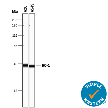
Detection of Human and Mouse HO‑1/HMOX1/HSP32 by Simple WesternTM. Simple Western lane view shows lysates of A20 mouse B cell lymphoma cell line and A549 human lung carcinoma cell line, loaded at 0.2 mg/mL. A specific band was detected for HO-1/HMOX1/HSP32 at approximately 37 kDa (as indicated) using 25 µg/mL of Goat Anti-Human/Mouse HO-1/HMOX1/HSP32 Antigen Affinity-purified Polyclonal Antibody (Catalog # AF3776) followed by 1:50 dilution of HRP-conjugated Anti-Goat IgG Secondary Antibody (Catalog # HAF109). This experiment was conducted under reducing conditions and using the 12-230 kDa separation system.
 View Larger
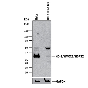
Western Blot Shows Human HO‑1/HMOX1/HSP32 Specificity by Using Knockout Cell Line. Western blot shows lysates of HeLa human cervical epithelial carcinoma parental cell line and HO-1/HMOX1/HSP32 knockout HeLa cell line (KO). PVDF membrane was probed with 0.5 µg/mL of Goat Anti-Human/Mouse HO-1/HMOX1/HSP32 Antigen Affinity-purified Polyclonal Antibody (Catalog # AF3776) followed by HRP-conjugated Anti-Goat IgG Secondary Antibody (Catalog # HAF017). A specific band was detected for HO-1/HMOX1/HSP32 at approximately 32 kDa (as indicated) in the parental HeLa cell line, but is not detectable in knockout HeLa cell line. GAPDH (Catalog # AF5718) is shown as a loading control. This experiment was conducted under reducing conditions and using Immunoblot Buffer Group 1.
Reconstitution Calculator
Preparation and Storage
- 12 months from date of receipt, -20 to -70 °C as supplied.
- 1 month, 2 to 8 °C under sterile conditions after reconstitution.
- 6 months, -20 to -70 °C under sterile conditions after reconstitution.
Background: HO-1/HMOX1/HSP32
Heme Oxygenase 1 (HO-1), also known as HMOX1 and Heat Shock Protein 32 (HSP32), is a 32 kDa microsomal enzyme required for the metabolism of heme to biliverdin. Heme oxygenase occurs as 2 isozymes, an inducible heme oxygenase-1 (HO-1/HMOX1) and a constitutive heme oxygenase-2 (HO-2/HMOX2). HO-1 expression is induced by heme and other non-heme compounds. Human HO-1 shares 82% amino acid sequence identity with mouse HO-1.
Product Datasheets
Citations for Human/Mouse HO-1/HMOX1/HSP32 Antibody
R&D Systems personnel manually curate a database that contains references using R&D Systems products. The data collected includes not only links to publications in PubMed, but also provides information about sample types, species, and experimental conditions.
3
Citations: Showing 1 - 3
Filter your results:
Filter by:
-
Allyl Isothiocyanate Protects Acetaminophen-Induced Liver Injury via NRF2 Activation by Decreasing Spontaneous Degradation in Hepatocyte
Authors: MW Kim, JH Kang, HJ Jung, SY Park, THL Phan, H Namgung, SY Seo, YS Yoon, SH Oh
Nutrients, 2020-11-23;12(11):.
Species: Mouse
Sample Types: Protein
Applications: Western Blot -
Red Yeast Rice Protects Circulating Bone Marrow-Derived Proangiogenic Cells against High-Glucose-Induced Senescence and Oxidative Stress: The Role of Heme Oxygenase-1
Authors: JT Liu, HY Chen, WC Chen, KM Man, YH Chen
Oxid Med Cell Longev, 2017-05-06;2017(0):3831750.
Species: Human
Sample Types: Cell Lysates
Applications: Western Blot -
Anti-inflammatory effects of ethyl acetate fraction from Melilotus suaveolens Ledeb on LPS-stimulated RAW 264.7 cells.
Authors: Tao JY, Zheng GH, Zhao L, Wu JG, Zhang XY, Zhang SL, Huang ZJ, Xiong FL, Li CM
J Ethnopharmacol, 2009-03-03;123(1):97-105.
Species: Mouse
Sample Types: Cell Lysates
Applications: Western Blot
FAQs
No product specific FAQs exist for this product, however you may
View all Antibody FAQsReviews for Human/Mouse HO-1/HMOX1/HSP32 Antibody
There are currently no reviews for this product. Be the first to review Human/Mouse HO-1/HMOX1/HSP32 Antibody and earn rewards!
Have you used Human/Mouse HO-1/HMOX1/HSP32 Antibody?
Submit a review and receive an Amazon gift card.
$25/€18/£15/$25CAN/¥75 Yuan/¥2500 Yen for a review with an image
$10/€7/£6/$10 CAD/¥70 Yuan/¥1110 Yen for a review without an image

