Human/Mouse Lipoprotein Lipase/LPL Antibody Summary
Applications
Please Note: Optimal dilutions should be determined by each laboratory for each application. General Protocols are available in the Technical Information section on our website.
Scientific Data
 View Larger
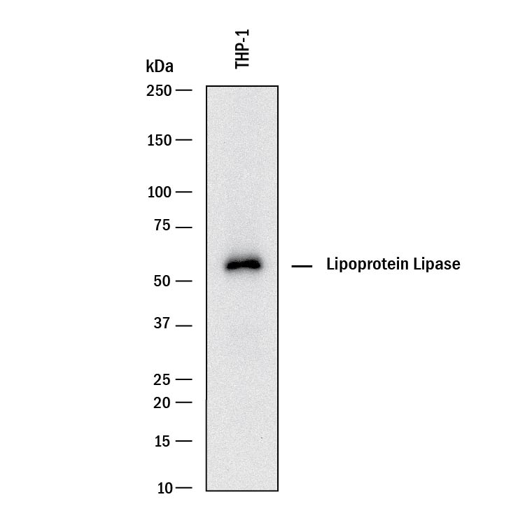
Detection of Human Lipoprotein Lipase/LPL by Western Blot. Western blot shows lysates of THP-1 human acute monocytic leukemia cell line. PVDF membrane was probed with 1 µg/mL of Goat Anti-Human/Mouse Lipoprotein Lipase/LPL Antigen Affinity-purified Polyclonal Antibody (Catalog # AF7197) followed by HRP-conjugated Anti-Goat IgG Secondary Antibody (Catalog # HAF017). A specific band was detected for Lipoprotein Lipase/LPL at approximately 55 kDa (as indicated). This experiment was conducted under reducing conditions and using Immunoblot Buffer Group 1.
 View Larger
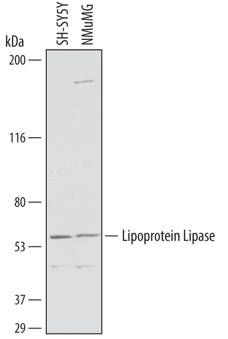
Detection of Human and Mouse Lipoprotein Lipase/LPL by Western Blot. Western blot shows lysates of SH-SY5Y human neuroblastoma cell line and NMuMG mouse mammary gland epithelial cell line. PVDF membrane was probed with 1 µg/mL of Goat Anti-Human Lipoprotein Lipase/LPL Antigen Affinity-purified Polyclonal Antibody (Catalog # AF7197) followed by HRP-conjugated Anti-Goat IgG Secondary Antibody (Catalog # HAF019). A specific band was detected for Lipoprotein Lipase/LPL at approximately 56 kDa (as indicated). This experiment was conducted under reducing conditions and using Immunoblot Buffer Group 1.
 View Larger
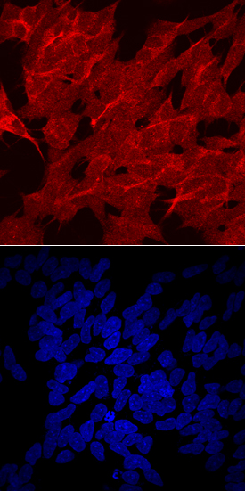
Lipoprotein Lipase/LPL in SH‑SY5Y Human Cell Line. Lipoprotein Lipase/LPL was detected in immersion fixed SH-SY5Y human neuroblastoma cell line using Goat Anti-Human Lipoprotein Lipase/LPL Antigen Affinity-purified Polyclonal Antibody (Catalog # AF7197) at 10 µg/mL for 3 hours at room temperature. Cells were stained using the NorthernLights™ 557-conjugated Anti-Goat IgG Secondary Antibody (red, upper panel; Catalog # NL001) and counterstained with DAPI (blue, lower panel). Specific staining was localized to cell surfaces and cytoplasm. View our protocol for Fluorescent ICC Staining of Cells on Coverslips. This application has not been tested in mouse samples.
 View Larger
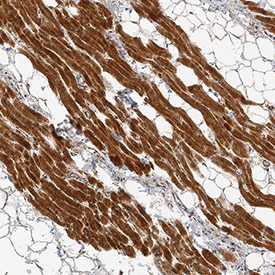
Lipoprotein Lipase/LPL in Human Heart. Lipoprotein Lipase/LPL was detected in immersion fixed paraffin-embedded sections of human heart using Goat Anti-Human/Mouse Lipoprotein Lipase/LPL Antigen Affinity-purified Polyclonal Antibody (Catalog # AF7197) at 3 µg/mL for 1 hour at room temperature followed by incubation with the Anti-Goat IgG VisUCyte™ HRP Polymer Antibody (Catalog # VC004). Tissue was stained using DAB (brown) and counterstained with hematoxylin (blue). Specific staining was localized to cardiomyocytes. View our protocol for IHC Staining with VisUCyte HRP Polymer Detection Reagents.
Reconstitution Calculator
Preparation and Storage
- 12 months from date of receipt, -20 to -70 degreesC as supplied. 1 month, 2 to 8 degreesC under sterile conditions after reconstitution. 6 months, -20 to -70 degreesC under sterile conditions after reconstitution.
Background: Lipoprotein Lipase/LPL
LPL (LipoProtein Lipase; also LIPD) is a 53-56 kDa glycoprotein member of the Lipase family, AB Hydrolase superfamily of molecules. It is produced by multiple cell types, including adipocytes, skelelal muscle cells and macrophages. Once secreted, the circulating enzyme ultimately becomes immobilized on the surface of endothelium by binding to cell surface heparan sulfate. Here, it hydrolyzes triglycerides embedded in chylomicrons and VLDLs by homodimerizing and interacting with apoC2. Mature human LPL is 448 amino acids (aa) in length. It contains an enzymatic region (aa 37-334) plus one protein-interaction PLAT domain (aa 341-465). Over aa 28-154, human LPL shares 91% aa identity with mouse LPL.
Product Datasheets
Citations for Human/Mouse Lipoprotein Lipase/LPL Antibody
R&D Systems personnel manually curate a database that contains references using R&D Systems products. The data collected includes not only links to publications in PubMed, but also provides information about sample types, species, and experimental conditions.
3
Citations: Showing 1 - 3
Filter your results:
Filter by:
-
The mechanisms to dispose of misfolded proteins in the endoplasmic reticulum of adipocytes
Authors: Wu, SA;Shen, C;Wei, X;Zhang, X;Wang, S;Chen, X;Torres, M;Lu, Y;Lin, LL;Wang, HH;Hunter, AH;Fang, D;Sun, S;Ivanova, MI;Lin, Y;Qi, L;
Nature communications
Species: Mouse, Transgenic Mouse
Sample Types: Whole Tissue, Cell Lysates
Applications: Immunohistochemistry, Immunoprecipitation, Immunogold Electron Microscopy, Western Blot -
Characterization of lipoprotein lipase storage vesicles in 3T3-L1 adipocytes
Authors: B Roberts, C Yang, S Neher
Journal of Cell Science, 2021-01-01;0(0):.
Species: Mouse
Sample Types: Cell Lysates, Whole Cells
Applications: ICC, Western Blot -
Firemaster� 550 and its components isopropylated triphenyl phosphate and triphenyl phosphate enhance adipogenesis and transcriptional activity of peroxisome proliferator activated receptor (Ppar?) on the adipocyte protein 2 (aP2) promoter
Authors: EWY Tung, S Ahmed, V Peshdary, E Atlas
PLoS ONE, 2017-04-24;12(4):e0175855.
Species: Mouse
Sample Types: Cell Lysates
Applications: Western Blot
FAQs
No product specific FAQs exist for this product, however you may
View all Antibody FAQsReviews for Human/Mouse Lipoprotein Lipase/LPL Antibody
There are currently no reviews for this product. Be the first to review Human/Mouse Lipoprotein Lipase/LPL Antibody and earn rewards!
Have you used Human/Mouse Lipoprotein Lipase/LPL Antibody?
Submit a review and receive an Amazon gift card.
$25/€18/£15/$25CAN/¥75 Yuan/¥2500 Yen for a review with an image
$10/€7/£6/$10 CAD/¥70 Yuan/¥1110 Yen for a review without an image


