Human/Mouse/Rat Annexin A1 Antibody Summary
Met1-Asn346
Accession # P04083
Applications
Please Note: Optimal dilutions should be determined by each laboratory for each application. General Protocols are available in the Technical Information section on our website.
Scientific Data
 View Larger
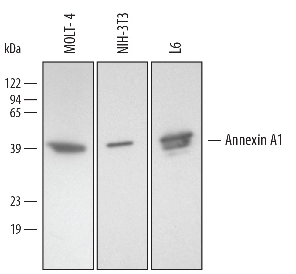
Detection of Human/Mouse/Rat Annexin A1 by Western Blot. Western blot shows lysates of MOLT-4 human acute lymphoblastic leukemia cell line, NIH-3T3 mouse embryonic fibroblast cell line, and L6 rat myoblast cell line. PVDF membrane was probed with 0.2 µg/mL of Goat Anti-Human/Mouse/Rat Annexin A1 Antigen Affinity-purified Polyclonal Antibody (Catalog # AF3770) followed by HRP-conjugated Anti-Goat IgG Secondary Antibody (Catalog # HAF109). A specific band was detected for Annexin A1 at approximately 35-40 kDa (as indicated). This experiment was conducted under reducing conditions and using Immunoblot Buffer Group 2.
 View Larger
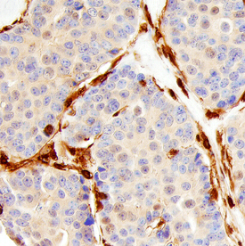
Annexin A1/Annexin I in Human Breast. Annexin A1/Annexin I was detected in immersion fixed paraffin-embedded sections of human breast using Goat Anti-Human/Mouse/Rat Annexin A1/Annexin I Antigen Affinity-purified Polyclonal Antibody (Catalog # AF3770) at 10 µg/mL overnight at 4 °C. Before incubation with the primary antibody, tissue was subjected to heat-induced epitope retrieval using Antigen Retrieval Reagent-Basic (Catalog # CTS013). Tissue was stained using the Anti-Goat HRP-DAB Cell & Tissue Staining Kit (brown; Catalog # CTS008) and counter-stained with hematoxylin (blue). Specific staining was localized to stromal cells. View our protocol for Chromogenic IHC Staining of Paraffin-embedded Tissue Sections.
 View Larger
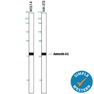
Detection of Human and Mouse Annexin A1 by Simple WesternTM. Simple Western lane view shows lysates of MOLT-4 human acute lymphoblastic leukemia cell line and NIH-3T3 mouse embryonic fibroblast cell line, loaded at 0.2 mg/mL. A specific band was detected for Annexin A1 at approximately 44 kDa (as indicated) using 2 µg/mL of Goat Anti-Human/Mouse/Rat Annexin A1 Antigen Affinity-purified Polyclonal Antibody (Catalog # AF3770) followed by 1:50 dilution of HRP-conjugated Anti-Goat IgG Secondary Antibody (Catalog # HAF109). This experiment was conducted under reducing conditions and using the 12-230 kDa separation system.
Reconstitution Calculator
Preparation and Storage
- 12 months from date of receipt, -20 to -70 °C as supplied.
- 1 month, 2 to 8 °C under sterile conditions after reconstitution.
- 6 months, -20 to -70 °C under sterile conditions after reconstitution.
Background: Annexin A1
The Annexins are a family of Calcium-dependent phospholipid-binding proteins that are preferentially located on the cytosolic face of the plasma membrane. The Annexin’s have a molecular weight of approximately 35 to 40 kDa and consist of a unique amino terminal domain followed by a homologous C-terminal core domain containing the calcium-dependent phospholipid-binding sites. The C‑terminal domain is comprised of four 60‑70 amino acid repeats, known as annexin repeats or an endonexin fold (Annexin A6 contains 8 annexin repeats). The four annexin repeats form a highly alpha -helical, tightly packed disc known as the annexin domain, which binds to phospholipids in the membrane in a calcium-dependent manner. Members of the annexin family play a role in cytoskeletal interactions, phospholipase inhibition, regulation of cellular growth, and intracellular signal transduction pathways. Annexin A1 (ANXA1), also known as annexin I, lipocortin I, and calpactin II, is an ~ 40 kDa protein with phospholipase A2 inhibitory activity. Since phospholipase A2 is required for the biosynthesis of the potent mediators of inflammation, prostaglandins and leukotrienes, Annexin A1 may have anti-inflammatory activity. Human Annexin A1 shares 88 and 89% identity with mouse and rat Annexin A1, respectively.
Product Datasheets
Citations for Human/Mouse/Rat Annexin A1 Antibody
R&D Systems personnel manually curate a database that contains references using R&D Systems products. The data collected includes not only links to publications in PubMed, but also provides information about sample types, species, and experimental conditions.
3
Citations: Showing 1 - 3
Filter your results:
Filter by:
-
Secreted KIAA1199 promotes the progression of rheumatoid arthritis by mediating hyaluronic acid degradation in an ANXA1-dependent manner
Authors: W Zhang, G Yin, H Zhao, H Ling, Z Xie, C Xiao, Y Chen, Y Lin, T Jiang, S Jin, J Wang, X Yang
Cell Death & Disease, 2021-01-20;12(1):102.
Species: Human
Sample Types: Whole Tissue
Applications: IHC -
Phenotypic and Expressional Heterogeneity in the Invasive Glioma Cells
Authors: A Fayzullin, CJ Sandberg, M Spreadbury, BM Saberniak, Z Grieg, E Skaga, IA Langmoen, EO Vik-Mo
Transl Oncol, 2018-10-03;12(1):122-133.
Species: Human
Sample Types: Whole Cells
Applications: ICC -
A role for stroma-derived annexin A1 as mediator in the control of genetic susceptibility to T-cell lymphoblastic malignancies through prostaglandin E2 secretion.
Authors: Santos J, Gonzalez-Sanchez L, Matabuena-Deyzaguirre M, Villa-Morales M, Cozar P, Lopez-Nieva P, Fernandez-Navarro P, Fresno M, Diaz-Munoz MD, Guenet JL, Montagutelli X, Fernandez-Piqueras J
Cancer Res., 2009-02-24;69(6):2577-87.
Species: Mouse
Sample Types: Cell Lysates
Applications: Western Blot
FAQs
No product specific FAQs exist for this product, however you may
View all Antibody FAQsReviews for Human/Mouse/Rat Annexin A1 Antibody
Average Rating: 4 (Based on 1 Review)
Have you used Human/Mouse/Rat Annexin A1 Antibody?
Submit a review and receive an Amazon gift card.
$25/€18/£15/$25CAN/¥75 Yuan/¥2500 Yen for a review with an image
$10/€7/£6/$10 CAD/¥70 Yuan/¥1110 Yen for a review without an image
Filter by:


