Human/Mouse/Rat DEP-1/CD148 Antibody Summary
Arg997-Ala1337
Accession # Q12913
Applications
Please Note: Optimal dilutions should be determined by each laboratory for each application. General Protocols are available in the Technical Information section on our website.
Scientific Data
 View Larger
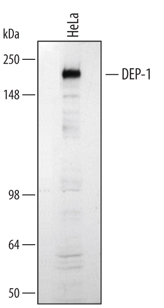
Detection of Human DEP‑1/CD148 by Western Blot. Western blot shows lysates of HeLa human cervical epithelial carcinoma cell line. PVDF membrane was probed with 1 µg/mL of Goat Anti-Human/Mouse/Rat DEP-1/CD148 Antigen Affinity-purified Polyclonal Antibody (Catalog # AF1934) followed by HRP-conjugated Anti-Goat IgG Secondary Antibody (Catalog # HAF109). A specific band was detected for DEP-1/CD148 at approximately 220 kDa (as indicated). This experiment was conducted under reducing conditions and using Immunoblot Buffer Group 1.
Reconstitution Calculator
Preparation and Storage
- 12 months from date of receipt, -20 to -70 °C as supplied.
- 1 month, 2 to 8 °C under sterile conditions after reconstitution.
- 6 months, -20 to -70 °C under sterile conditions after reconstitution.
Background: DEP-1/CD148
Density Enhanced Protein Tyrosine Phosphatase (DEP-1), also known as CD148, HPTP-eta, and PTP receptor type J (PTPRJ), is an enzyme that removes phosphate groups covalently attached to tyrosine residues in proteins. A large (220 kilodalton) glycoprotein found at the cell surface, DEP-1 levels are increased with high cell density (1). DEP-1 phosphatase activity is enhanced by basement membrane proteins (2), suggesting it is involved in regulating cell adhesion and contact interactions. High levels of expression dampen PDGF (3), VEGF (4), and T-cell receptor (5) responses. DEP-1 is widely expressed in tissues, particularly ones forming epithelioid monolayers (6). In the immune system, DEP-1 is found on all cell lineages and is highest on granulocytes (7). Dep-1 is the mutated gene in the Susceptibility to Colon Cancer locus Scc1, which is altered in many human colorectal adenomas (8). Gene knockout mice lacking DEP-1 die at midgestation due to failures in cardiovascular development (9). DEP-1 dephosphorylates a variety of proteins, including the HGF (10), PDGF (11), and VEGF (4) receptors, and beta ‑catenin (12). The recombinant protein is the intracellular region of DEP-1 containing the catalytic domain. Over aa 997-337, human Dep-1 shares 95% aa sequence identity with mouse and rat Dep-1.
- Ostman, A. et al. (1994) Proc. Natl. Acad. Sci. USA 91:9680.
- Sorby, M. et al. (2001) Oncogene 20:5219.
- Jandt, E. et al. (2003) Oncogene 22:4175.
- Lampugnani, M.G. et al. (2003) J. Cell Biol. 161:793.
- Baker, J.E. et al. (2001) Mol. Cell. Biol. 21:2393.
- Borges, L.G. et al. (1996) Circ. Res. 79:570.
- de la Fuente-Garcia, M.A. et al. (1998) Blood 91:2800.
- Ruivenkamp, C.A. et al. (2002) Nat. Genet. 31:295.
- Takahashi, T. et al. (2003) Mol. Cell. Biol. 23:1817.
- Palka, H.L. et al. (2003) J. Biol. Chem. 278:5728.
- Kovalenko, M. et al. (2000) J. Biol. Chem. 275:16219.
- Holsinger, L.J. et al. (2002) Oncogene 21:7067.
Product Datasheets
Citations for Human/Mouse/Rat DEP-1/CD148 Antibody
R&D Systems personnel manually curate a database that contains references using R&D Systems products. The data collected includes not only links to publications in PubMed, but also provides information about sample types, species, and experimental conditions.
4
Citations: Showing 1 - 4
Filter your results:
Filter by:
-
PTPRJ Inhibits Leptin Signaling, and Induction of PTPRJ in the Hypothalamus Is a Cause of the Development of Leptin Resistance
Authors: T Shintani, S Higashi, R Suzuki, Y Takeuchi, R Ikaga, T Yamazaki, K Kobayashi, M Noda
Sci Rep, 2017-09-14;7(1):11627.
Species: Human, Mouse
Sample Types: Cell Lysates, Whole Cells
Applications: ICC, Western Blot -
Phosphorylation of DEP-1/PTPRJ on threonine 1318 regulates Src activation and endothelial cell permeability induced by vascular endothelial growth factor.
Authors: Spring K, Lapointe L, Caron C, Langlois S, Royal I
Cell Signal, 2014-02-28;26(6):1283-93.
Species: Human
Sample Types: Recombinant Protein
Applications: Immunoprecipitation -
T cell Ig and mucin domain-containing protein 3 is recruited to the immune synapse, disrupts stable synapse formation, and associates with receptor phosphatases.
Authors: Clayton K, Haaland M, Douglas-Vail M, Mujib S, Chew G, Ndhlovu L, Ostrowski M
J Immunol, 2013-12-13;192(2):782-91.
Species: Human
Sample Types: Cell Lysates
Applications: Western Blot -
Protein-tyrosine phosphatase DEP-1 controls receptor tyrosine kinase FLT3 signaling.
Authors: Arora D, Stopp S, Bohmer SA, Schons J, Godfrey R, Masson K, Razumovskaya E, Ronnstrand L, Tanzer S, Bauer R, Bohmer FD, Muller JP
J. Biol. Chem., 2011-01-24;286(13):10918-29.
Species: Human
Sample Types: Whole Cells
Applications: Flow Cytometry
FAQs
No product specific FAQs exist for this product, however you may
View all Antibody FAQsReviews for Human/Mouse/Rat DEP-1/CD148 Antibody
There are currently no reviews for this product. Be the first to review Human/Mouse/Rat DEP-1/CD148 Antibody and earn rewards!
Have you used Human/Mouse/Rat DEP-1/CD148 Antibody?
Submit a review and receive an Amazon gift card.
$25/€18/£15/$25CAN/¥75 Yuan/¥2500 Yen for a review with an image
$10/€7/£6/$10 CAD/¥70 Yuan/¥1110 Yen for a review without an image

