Human/Mouse/Rat FABP5/E-FABP Antibody Summary
Met1-Gln135
Accession # Q05816
Applications
Please Note: Optimal dilutions should be determined by each laboratory for each application. General Protocols are available in the Technical Information section on our website.
Scientific Data
 View Larger
Detection of Human, Mouse, and Rat FABP5/E‑FABP by Western Blot. Western blot shows lysates of human placenta tissue, mouse liver tissue, rat brain tissue, and rat heart tissue. PVDF membrane was probed with 0.5 µg/mL of Goat Anti-Human/Mouse/ Rat FABP5/E-FABP Antigen Affinity-purified Polyclonal Antibody (Catalog # AF1476) followed by HRP-conjugated Anti-Goat IgG Secondary Antibody (HAF017). A specific band was detected for FABP5/ E-FABP at approximately 15 kDa (as indicated). This experiment was conducted under reducing conditions and using Immunoblot Buffer Group 1.
.") View Larger
View Larger
FABP5/E‑FABP in Mouse Skin Tissue. FABP5/E-FABP was detected in immersion fixed paraffin-embedded sections of mouse skin (ear) tissue using Goat Anti-Human/Mouse/Rat FABP5/E-FABP Antigen Affinity-purified Polyclonal Antibody (Catalog # AF1476) at 3 µg/mL for 1 hour at room temperature followed by incubation with the Anti-Goat IgG VisUCyte™ HRP Polymer Antibody (VC004). Before incubation with the primary antibody, tissue was subjected to heat-induced epitope retrieval using Antigen Retrieval Reagent-Basic (CTS013). Tissue was stained using DAB (brown) and counterstained with hematoxylin (blue). Specific staining was localized to cell nuclei. View our protocol for IHC Staining with VisUCyte HRP Polymer Detection Reagents.
 View Larger
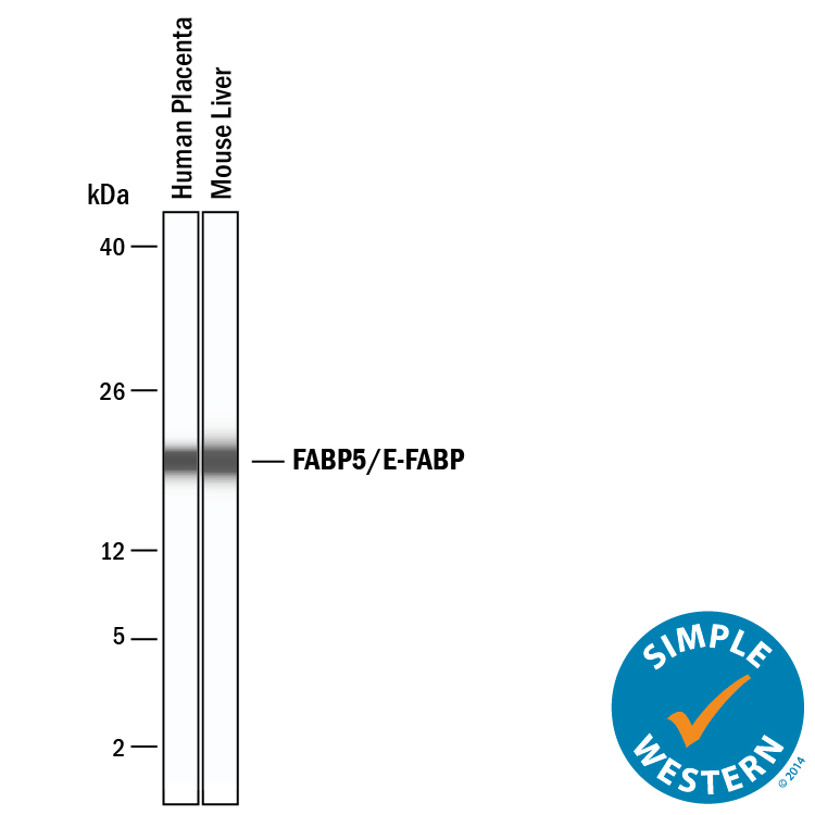
Detection of Human and Mouse FABP5/E‑FABP by Simple WesternTM. Simple Western lane view shows lysates of human placenta and mouse liver, loaded at 0.2 mg/mL. A specific band was detected for FABP5/E‑FABP at approximately 20 kDa (as indicated) using 10 µg/mL of Goat Anti-Human/Mouse/Rat FABP5/E‑FABP Antigen Affinity-purified Polyclonal Antibody (Catalog # AF1476). This experiment was conducted under reducing conditions and using the 2-40 kDa separation system.
 View Larger
Western Blot Shows Human FABP5/E‑FABP Specificity by Using Knockout Cell Line. Western blot shows lysates of HeLa human cervical epithelial carcinoma parental cell line and FABP5/E-FABP knockout HeLa cell line (KO). PVDF membrane was probed with 0.5 µg/mL of Goat Anti-Human/Mouse/Rat FABP5/E-FABP Antigen Affinity-purified Polyclonal Antibody (Catalog # AF1476) followed by HRP-conjugated Anti-Goat IgG Secondary Antibody (HAF017). A specific band was detected for FABP5/E-FABP at approximately 15 kDa (as indicated) in the parental HeLa cell line, but is not detectable in knockout HeLa cell line. GAPDH (AF5718) is shown as a loading control. This experiment was conducted under reducing conditions and using Immunoblot Buffer Group 1.
Reconstitution Calculator
Preparation and Storage
- 12 months from date of receipt, -20 to -70 °C as supplied.
- 1 month, 2 to 8 °C under sterile conditions after reconstitution.
- 6 months, -20 to -70 °C under sterile conditions after reconstitution.
Background: FABP5/E-FABP
FABP5, also known as epidermal fatty acid binding protein (E-FABP), is a 15 kDa member of a cytosolic fatty acid binding protein superfamily. It is associated with keratinocytes and adipocytes and is suggested to promote fatty acid availability to enzymes, protect cell structures from fatty acid attack, and target fatty acids to nuclear transcription factors. The amino acid sequence of human FABP5 is 80%, 81% and 92% identical to that of mouse, rat and bovine FABP5, respectively.
Product Datasheets
Citations for Human/Mouse/Rat FABP5/E-FABP Antibody
R&D Systems personnel manually curate a database that contains references using R&D Systems products. The data collected includes not only links to publications in PubMed, but also provides information about sample types, species, and experimental conditions.
14
Citations: Showing 1 - 10
Filter your results:
Filter by:
-
Genetic or pharmacologic blockade of mPGES-2 attenuates renal lipotoxicity and diabetic kidney disease by targeting Rev-Erb?/FABP5 signaling
Authors: Zhong, D;Chen, J;Qiao, R;Song, C;Hao, C;Zou, Y;Bai, M;Su, W;Yang, B;Sun, D;Jia, Z;Sun, Y;
Cell reports
Species: Transgenic Mouse, Mouse
Sample Types: Whole Tissue
Applications: Immunohistochemistry -
Fatty Acid-Binding Protein 5 Modulates Brain Endocannabinoid Tone and Retrograde Signaling in the Striatum
Authors: M Fauzan, S Oubraim, M Yu, ST Glaser, M Kaczocha, S Haj-Dahman
Frontiers in Cellular Neuroscience, 2022-07-07;16(0):936939.
Species: Mouse
Sample Types: Whole Tissue
Applications: IHC -
FABP5 deletion in nociceptors augments endocannabinoid signaling and suppresses TRPV1 sensitization and inflammatory pain
Authors: DM Bogdan, K Studholme, A DiBua, C Gordon, MP Kanjiya, M Yu, M Puopolo, M Kaczocha
Oncogene, 2022-06-02;12(1):9241.
Species: Mouse
Sample Types: Whole Tissue
Applications: IHC -
Stroke induces disease-specific myeloid cells in the brain parenchyma and pia
Authors: C Beuker, D Schafflick, JK Strecker, M Heming, X Li, J Wolbert, A Schmidt-Po, C Thomas, T Kuhlmann, I Aranda-Par, N A-Gonzalez, PA Kumar, Y Werner, E Kilic, DM Hermann, H Wiendl, R Stumm, GMZ Hörste, J Minnerup
Nature Communications, 2022-02-17;13(1):945.
Species: Human, Mouse
Sample Types: Whole Tissue
Applications: IHC -
Destructing biofilms by cationic dextran through phase transition
Authors: Y Li, S Wang, Z Xing, Y Niu, Z Liao, Y Lu, J Qiu, J Zhang, C Wang, L Dong
Carbohydrate polymers, 2021-10-23;279(0):118778.
Species: Mouse
Sample Types: Whole Tissue
Applications: IHC -
JunB defines functional and structural integrity of the epidermo-pilosebaceous unit in the skin
Authors: K Singh, E Camera, L Krug, A Basu, RK Pandey, S Munir, M Wlaschek, S Kochanek, M Schorpp-Ki, M Picardo, P Angel, C Niemann, P Maity, K Scharffett
Nat Commun, 2018-08-24;9(1):3425.
Species: Mouse
Sample Types: Whole Tissue
Applications: IHC-P -
Adipose Fatty Acid Binding Protein Promotes Saturated Fatty Acid-Induced Macrophage Cell Death through Enhancing Ceramide Production
Authors: Yuwen Zhang
J. Immunol, 2016-12-05;0(0):.
Species: Mouse
Sample Types: Cell Lysates
Applications: Western Blot -
BLIMP1 is required for postnatal epidermal homeostasis but does not define a sebaceous gland progenitor under steady-state conditions.
Authors: Kretzschmar K, Cottle D, Donati G, Chiang M, Quist S, Gollnick H, Natsuga K, Lin K, Watt F
Stem Cell Reports, 2014-09-18;3(4):620-33.
Species: Rat
Sample Types: Whole Tissue
Applications: IHC -
Fatty acid-binding protein E-FABP restricts tumor growth by promoting IFN-beta responses in tumor-associated macrophages.
Authors: Zhang Y, Sun Y, Rao E, Yan F, Li Q, Zhang Y, Silverstein K, Liu S, Sauter E, Cleary M, Li B
Cancer Res, 2014-04-08;74(11):2986-98.
Species: Mouse
Sample Types: Cell Lysates, Whole Cells, Whole Tissue
Applications: IHC, Western Blot -
Fatty acid-binding protein 5 (FABP5) regulates cognitive function both by decreasing anandamide levels and by activating the nuclear receptor peroxisome proliferator-activated receptor beta/delta (PPARbeta/delta) in the brain.
Authors: Yu S, Levi L, Casadesus G, Kunos G, Noy N
J Biol Chem, 2014-03-18;289(18):12748-58.
Species: Mouse
Sample Types: Cell Lysates
Applications: Western Blot -
Retinoic acid induces neurogenesis by activating both retinoic acid receptors (RARs) and peroxisome proliferator-activated receptor beta/delta (PPARbeta/delta).
Authors: Yu S, Levi L, Siegel R, Noy N
J Biol Chem, 2012-10-26;287(50):42195-205.
Species: Mouse
Sample Types: Cell Lysates, Whole Cells, Whole Tissue
Applications: ICC, IHC-Fr, Western Blot -
Proteomic analysis of proteins differentially expressed in uterine lymphocytes obtained from wild-type and NOD mice.
Authors: Li C, Wang W, Wang H, Zhong Y, Di J, Lin Y
J. Cell. Biochem., 2009-10-01;108(2):447-57.
Species: Mouse
Sample Types: Cell Lysates
Applications: Western Blot -
All-trans-retinoic acid represses obesity and insulin resistance by activating both peroxisome proliferation-activated receptor beta/delta and retinoic acid receptor.
Authors: Berry DC, Noy N
Mol. Cell. Biol., 2009-04-13;29(12):3286-96.
Species: Mouse
Sample Types: Cell Lysates
Applications: Western Blot -
Helper-dependent adenovirus-mediated short hairpin RNA expression in the liver activates the interferon response.
Authors: Witting SR, Brown M, Saxena R, Nabinger S, Morral N
J. Biol. Chem., 2007-11-19;283(4):2120-8.
Species: Mouse
Sample Types: Tissue Homogenates
Applications: Western Blot
FAQs
No product specific FAQs exist for this product, however you may
View all Antibody FAQsReviews for Human/Mouse/Rat FABP5/E-FABP Antibody
There are currently no reviews for this product. Be the first to review Human/Mouse/Rat FABP5/E-FABP Antibody and earn rewards!
Have you used Human/Mouse/Rat FABP5/E-FABP Antibody?
Submit a review and receive an Amazon gift card.
$25/€18/£15/$25CAN/¥75 Yuan/¥2500 Yen for a review with an image
$10/€7/£6/$10 CAD/¥70 Yuan/¥1110 Yen for a review without an image

