Human/Mouse/Rat Glutathione Peroxidase 1/GPX1 Antibody
Human/Mouse/Rat Glutathione Peroxidase 1/GPX1 Antibody Summary
Gly48-Ala201
Accession # P07203
Applications
Please Note: Optimal dilutions should be determined by each laboratory for each application. General Protocols are available in the Technical Information section on our website.
Scientific Data
 View Larger
Glutathione Peroxidase 1/GPX1 in Rat Liver. Glutathione Peroxidase 1/GPX1 was detected in perfusion fixed frozen sections of rat liver using 15 µg/mL Goat Anti-Human/Mouse/Rat Glutathione Peroxidase 1/GPX1 Antigen Affinity-purified Polyclonal Antibody (Catalog # AF3798) overnight at 4 °C. Tissue was stained with the Anti-Goat HRP-DAB Cell & Tissue Staining Kit (brown; Catalog # CTS008) and counterstained with hematoxylin (blue). Specific labeling was localized to the cytoplasm in hepatocytes. View our protocol for Chromogenic IHC Staining of Frozen Tissue Sections.
 View Larger
Detection of Human/Mouse/Rat Glutathione Peroxidase 1 by Western Blot. Western blot shows lysates of human, mouse and rat liver tissue. PVDF membrane was probed with 1 µg/mL of Goat Anti-Human/Mouse/Rat Glutathione Peroxidase 1 Antigen Affinity-purified Polyclonal Antibody (Catalog # AF3798) followed by HRP-conjugated Anti-Goat IgG Secondary Antibody (Catalog # HAF017). A specific band was detected for Glutathione Peroxidase 1 at approximately 22 kDa (as indicated). This experiment was conducted under reducing conditions and using Immunoblot Buffer Group 2.
 View Larger
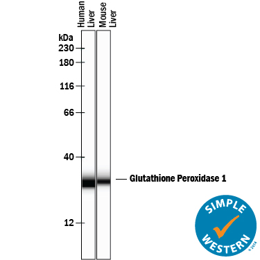
Detection of Human and Mouse Glutathione Peroxidase 1/GPX1 by Simple WesternTM. Simple Western lane view shows lysates of human liver tissue and mouse liver tissue, loaded at 0.2 mg/mL. A specific band was detected for Glutathione Peroxidase 1/GPX1 at approximately 30 kDa (as indicated) using 10 µg/mL of Goat Anti-Human/Mouse/Rat Glutathione Peroxidase 1/GPX1 Antigen Affinity-purified Polyclonal Antibody (Catalog # AF3798) followed by 1:50 dilution of HRP-conjugated Anti-Goat IgG Secondary Antibody (Catalog # HAF109). This experiment was conducted under reducing conditions and using the 12-230 kDa separation system.
 View Larger
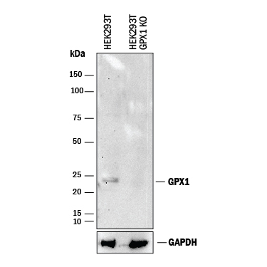
Western Blot Shows Human Glutathione Peroxidase 1/GPX1 Specificity by Using Knockout Cell Line. Western blot shows lysates of HEK293T human embryonic kidney parental cell line and Glutathione Peroxidase 1/GPX1 knockout HEK293T cell line (KO). PVDF membrane was probed with 1 µg/mL of Goat Anti-Human/Mouse/Rat Glutathione Peroxidase 1/GPX1 Antigen Affinity-purified Polyclonal Antibody (Catalog # AF3798) followed by HRP-conjugated Anti-Goat IgG Secondary Antibody (Catalog # HAF017). A specific band was detected for Glutathione Peroxidase 1/GPX1 at approximately 24 kDa (as indicated) in the parental HEK293T cell line, but is not detectable in knockout HEK293T cell line. GAPDH (Catalog # AF5718) is shown as a loading control. This experiment was conducted under reducing conditions and using Immunoblot Buffer Group 1.
Reconstitution Calculator
Preparation and Storage
- 12 months from date of receipt, -20 to -70 °C as supplied.
- 1 month, 2 to 8 °C under sterile conditions after reconstitution.
- 6 months, -20 to -70 °C under sterile conditions after reconstitution.
Background: Glutathione Peroxidase 1/GPX1
Glutathione Peroxidase 1 (GPX1), also known as Cellular Glutathione Peroxidase, is a 201 amino acid, 23 kDa member of the glutathione peroxidase antioxidant enzyme family. The Glutathione Peroxidase family protects cell surfaces, extracellular fluid components, and enzymes from oxidative stress by catalysing the reduction of hydrogen peroxide, lipid peroxides, and organic hydroperoxide using reduced glutathione. GPX1 is a 92 kDa homotetramer consisting of four identical 23 kDa subunits, each containing a selenocysteine residue at the active site. GPX1 is an important antioxidant enzyme and protects red blood cells and other cells from oxidative damage. Deficiencies in red cell GPX1 have been associated with hemolytic anemia. Two alternatively spliced transcript variants encoding distinct isoforms have been found for this gene.
Product Datasheets
Citations for Human/Mouse/Rat Glutathione Peroxidase 1/GPX1 Antibody
R&D Systems personnel manually curate a database that contains references using R&D Systems products. The data collected includes not only links to publications in PubMed, but also provides information about sample types, species, and experimental conditions.
16
Citations: Showing 1 - 10
Filter your results:
Filter by:
-
N-Acetylcysteine Reduces the Pro-Oxidant and Inflammatory Responses during Pancreatitis and Pancreas Tumorigenesis
Authors: Minati MA, Libert M, Dahou H et al.
Antioxidants (Basel)
-
Glutathione peroxidase-1 overexpression reduces oxidative stress, and improves pathology and proteome remodeling in the kidneys of old mice
Authors: Y Chu, RS Lan, R Huang, H Feng, R Kumar, S Dayal, KS Chan, DF Dai
Aging Cell, 2020-05-13;0(0):.
Species: Mouse
Sample Types: Tissue Homogenates
Applications: Western Blot -
Loss of epitranscriptomic control of selenocysteine utilization engages senescence and mitochondrial reprogramming?
Authors: MY Lee, A Leonardi, TJ Begley, JA Melendez
Redox Biol, 2019-11-11;28(0):101375.
Species: Mouse
Sample Types: Cell Lysates
Applications: Western Blot -
A Mitochondrial Specific Antioxidant Reverses Metabolic Dysfunction and Fatty Liver Induced by Maternal Cigarette Smoke in Mice
Authors: G Li, YL Chan, S Sukjamnong, AG Anwer, H Vindin, M Padula, R Zakarya, J George, BG Oliver, S Saad, H Chen
Nutrients, 2019-07-21;11(7):.
Species: Mouse
Sample Types: Protein
Applications: Western Blot -
Evidence supporting oxidative stress in a moderately affected area of the brain in Alzheimer's disease
Authors: P Youssef, B Chami, J Lim, T Middleton, GT Sutherland, PK Witting
Sci Rep, 2018-08-01;8(1):11553.
Species: Human
Sample Types: Tissue Homogenates
-
Dietary tetrahydrocurcumin reduces renal fibrosis and cardiac hypertrophy in 5/6 nephrectomized rats
Authors: WL Lau, M Khazaeli, J Savoj, K Manekia, M Bangash, RG Thakurta, A Dang, ND Vaziri, B Singh
Pharmacol Res Perspect, 2018-02-19;6(2):e00385.
Species: Rat
Sample Types: Tissue Homogenates
Applications: Western Blot -
Partial involvement of Nrf2 in skeletal muscle mitohormesis as an adaptive response to mitochondrial uncoupling
Authors: V Coleman, P Sa-Nguanmo, J Koenig, TJ Schulz, T Grune, S Klaus, AP Kipp, M Ost
Sci Rep, 2018-02-05;8(1):2446.
Species: Mouse
Sample Types: Tissue Homogenates
Applications: Western Blot -
Sexual Dimorphism in the Selenocysteine Lyase Knockout Mouse
Authors: AN Ogawa-Wong, AC Hashimoto, H Ha, MW Pitts, LA Seale, MJ Berry
Nutrients, 2018-01-31;10(2):.
Species: Mouse
Sample Types: Cell Lysate
Applications: Western Blot -
LCZ696 (Sacubitril/valsartan) ameliorates oxidative stress, inflammation, fibrosis and improves renal function beyond angiotensin receptor blockade in CKD
Authors: W Jing, ND Vaziri, A Nunes, Y Suematsu, T Farzaneh, M Khazaeli, H Moradi
Am J Transl Res, 2017-12-15;9(12):5473-5484.
Species: Rat
Sample Types: Tissue Homogenates
Applications: Western Blot -
Global survey of cell death mechanisms reveals metabolic regulation of ferroptosis
Nat Chem Biol, 2016-05-09;0(0):.
Species: Human
Sample Types: Cell Lysates
Applications: Western Blot -
Isoform-specific binding of selenoprotein P to the beta-propeller domain of apolipoprotein E receptor 2 mediates selenium supply.
Authors: Kurokawa S, Bellinger F, Hill K, Burk R, Berry M
J Biol Chem, 2014-02-13;289(13):9195-207.
Species: Human
Sample Types: Cell Lysates
Applications: Western Blot -
Mice lacking selenoprotein P and selenocysteine lyase exhibit severe neurological dysfunction, neurodegeneration, and audiogenic seizures.
Authors: Byrns, China N, Pitts, Matthew, Gilman, Christy, Hashimoto, Ann C, Berry, Marla J
J Biol Chem, 2014-02-11;289(14):9662-74.
Species: Mouse
Sample Types: Tissue Homogenates
Applications: Western Blot -
Evaluation of selenite effects on selenoproteins and cytokinome in human hepatoma cell lines.
Authors: Rusolo, Fabiola, Pucci, Biagio, Colonna, Giovanni, Capone, Francesc, Guerriero, Eliana, Milone, Maria Ri, Nazzaro, Melissa, Volpe, Maria Gr, Di Bernardo, Gianni, Castello, Giuseppe, Costantini, Susan
Molecules, 2013-02-26;18(3):2549-62.
Species: Human
Sample Types: Cell Lysates
Applications: Western Blot -
Prostaglandin H Synthase-2-catalyzed Oxygenation of 2-Arachidonoylglycerol Is More Sensitive to Peroxide Tone than Oxygenation of Arachidonic Acid.
Authors: Musee J, Marnett L
J Biol Chem, 2012-09-01;287(44):37383-94.
Species: Mouse
Sample Types: Cell Lysates
Applications: Western Blot -
Disruption of the selenocysteine lyase-mediated selenium recycling pathway leads to metabolic syndrome in mice.
Authors: Seale L, Hashimoto A, Kurokawa S, Gilman C, Seyedali A, Bellinger F, Raman A, Berry M
Mol Cell Biol, 2012-08-13;32(20):4141-54.
Species: Mouse
Sample Types: Tissue Homogenates
Applications: Western Blot -
Resveratrol reduces endothelial oxidative stress by modulating the gene expression of superoxide dismutase 1 (SOD1), glutathione peroxidase 1 (GPx1) and NADPH oxidase subunit (Nox4).
Authors: Spanier G, Xu H, Xia N
J. Physiol. Pharmacol., 2009-10-01;60(0):111-6.
Species: Human
Sample Types: Cell Lysates
Applications: Western Blot
FAQs
No product specific FAQs exist for this product, however you may
View all Antibody FAQsReviews for Human/Mouse/Rat Glutathione Peroxidase 1/GPX1 Antibody
There are currently no reviews for this product. Be the first to review Human/Mouse/Rat Glutathione Peroxidase 1/GPX1 Antibody and earn rewards!
Have you used Human/Mouse/Rat Glutathione Peroxidase 1/GPX1 Antibody?
Submit a review and receive an Amazon gift card.
$25/€18/£15/$25CAN/¥75 Yuan/¥2500 Yen for a review with an image
$10/€7/£6/$10 CAD/¥70 Yuan/¥1110 Yen for a review without an image

