Human/Mouse/Rat IGSF8/CD316 Antibody Summary
Ala25-Thr577
Accession # NP_536344
Applications
Please Note: Optimal dilutions should be determined by each laboratory for each application. General Protocols are available in the Technical Information section on our website.
Scientific Data
 View Larger
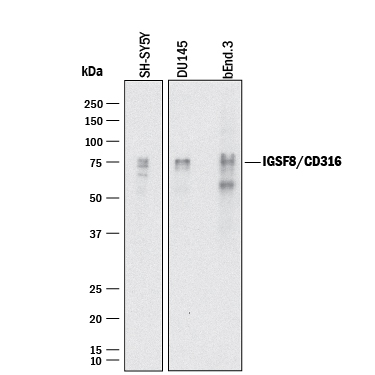
Detection of Human and Mouse IGSF8/CD316 by Western Blot. Western blot shows lysates of SH-SY5Y human neuroblastoma cell line, DU145 human prostate carcinoma cell line, and bEnd.3 mouse endothelioma cell line. PVDF membrane was probed with 1 µg/mL of Goat Anti-Human/Mouse/Rat IGSF8/CD316 Antigen Affinity-purified Polyclonal Antibody (Catalog # AF3117) followed by HRP-conjugated Anti-Goat IgG Secondary Antibody (Catalog # HAF017). Specific bands were detected for IGSF8/CD316 at approximately 70-80 kDa (as indicated). This experiment was conducted under reducing conditions and using Immunoblot Buffer Group 1.
 View Larger
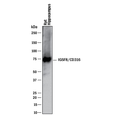
Detection of Rat IGSF8/CD316 by Western Blot. Western blot shows lysates of rat brain (hippocampus) tissue. PVDF membrane was probed with 1 µg/mL of Goat Anti-Human/Mouse/Rat IGSF8/CD316 Antigen Affinity-purified Polyclonal Antibody (Catalog # AF3117) followed by HRP-conjugated Anti-Goat IgG Secondary Antibody (Catalog # HAF017). A specific band was detected for IGSF8/CD316 at approximately 70 kDa (as indicated). This experiment was conducted under reducing conditions and using Immunoblot Buffer Group 1.
 View Larger
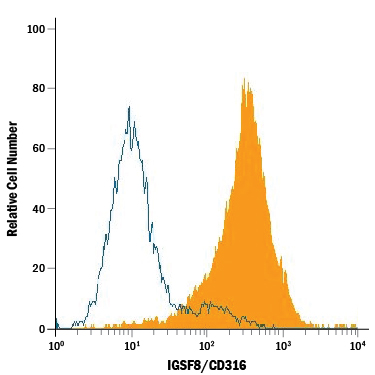
Detection of IGSF8/CD316 in Neuro‑2A Mouse Cell Line by Flow Cytometry. Neuro-2A mouse neuroblastoma cell line was stained with Goat Anti-Human/Mouse/Rat IGSF8/CD316 Antigen Affinity-purified Polyclonal Antibody (Catalog # AF3117, filled histogram) or isotype control antibody (Catalog # AB-108-C, open histogram), followed by Phycoerythrin-conjugated Anti-Goat IgG Secondary Antibody (Catalog # F0107).
 View Larger
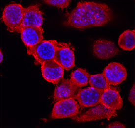
IGSF8/CD316 in Neuro‑2A Mouse Cell Line. IGSF8/CD316 was detected in immersion fixed Neuro-2A mouse neuroblastoma cell line using Goat Anti-Human/Mouse/Rat IGSF8/CD316 Antigen Affinity-purified Polyclonal Antibody (Catalog # AF3117) at 10 µg/mL for 3 hours at room temperature. Cells were stained using the NorthernLights™ 557-conjugated Anti-Goat IgG Secondary Antibody (red; Catalog # NL001) and counterstained with DAPI (blue). Specific staining was localized to cell surfaces. View our protocol for Fluorescent ICC Staining of Cells on Coverslips.
 View Larger
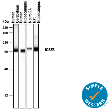
Detection of Human, Mouse, and Rat IGSF8/CD316 by Simple WesternTM. Simple Western lane view shows lysates of human cerebellum tissue, human hippocampus tissue, Neuro-2A mouse neuroblastoma cell line, and rat hippocampus tissue, loaded at 0.2 mg/mL. A specific band was detected for IGSF8/CD316 at approximately 71-80 kDa (as indicated) using 20 µg/mL of Goat Anti-Human/Mouse IGSF8/CD316 Antigen Affinity-purified Polyclonal Antibody (Catalog # AF3117) followed by 1:50 dilution of HRP-conjugated Anti-Goat IgG Secondary Antibody (Catalog # HAF109). This experiment was conducted under reducing conditions and using the 12-230 kDa separation system.
Reconstitution Calculator
Preparation and Storage
- 12 months from date of receipt, -20 to -70 °C as supplied.
- 1 month, 2 to 8 °C under sterile conditions after reconstitution.
- 6 months, -20 to -70 °C under sterile conditions after reconstitution.
Background: IGSF8/CD316
IGSF8, also known as PGRL (PG Regulatory-Like Protein), KASP (KAI/CD82 Associated Protein) and EWI-2 (Glu-Trp-Ile motif 2), is a widely expressed transmembrane adhesion protein. It interacts with beta 1 Integrins and various tetraspanins including CD9, CD81 and CD82. IGSF8 contains four extracellular Ig-like domains. IGSF8 over-expression in transformed cells inhibits cell migration and suppresses cancer metastatic potential. Mouse and human IGSF8 share 90% amino acid sequence identity.
Product Datasheets
Citations for Human/Mouse/Rat IGSF8/CD316 Antibody
R&D Systems personnel manually curate a database that contains references using R&D Systems products. The data collected includes not only links to publications in PubMed, but also provides information about sample types, species, and experimental conditions.
7
Citations: Showing 1 - 7
Filter your results:
Filter by:
-
MicroRNA-7 regulates melanocortin circuits involved in mammalian energy homeostasis
Authors: MP LaPierre, K Lawler, S Godbersen, IS Farooqi, M Stoffel
Nature Communications, 2022-09-29;13(1):5733.
Species: Mouse
Sample Types: Cell Lysates
Applications: Western Blot -
A conserved sequence in the small intracellular loop of tetraspanins forms an M-shaped inter-helix turn
Authors: N Reppert, T Lang
Scientific Reports, 2022-03-16;12(1):4494.
Species: Human
Sample Types: Cell Lysates
Applications: Western Blot -
Hspa8 and ICAM-1 as damage-induced mediators of &gamma&delta T cell activation
Authors: MD Johnson, MF Otuki, DA Cabrini, R Rudolph, DA Witherden, WL Havran
Journal of leukocyte biology, 2021-04-13;0(0):.
Species: Mouse
Sample Types: Whole Cells
Applications: Flow Cytometry -
Synapse type-specific proteomic dissection identifies IgSF8 as a hippocampal CA3 microcircuit organizer
Authors: N Apóstolo, SN Smukowski, J Vanderlind, G Condomitti, V Rybakin, J Ten Bos, L Trobiani, S Portegies, KM Vennekens, NV Gounko, D Comoletti, KD Wierda, JN Savas, J de Wit
Nat Commun, 2020-10-14;11(1):5171.
Species: Mouse, Rat
Sample Types: Cell Culture Supernates, Whole Tissue
Applications: ICC, IHC -
Transcriptome analysis reveals transmembrane targets on transplantable midbrain dopamine progenitors.
Authors: Bye C, Jonsson M, Bjorklund A, Parish C, Thompson L
Proc Natl Acad Sci U S A, 2015-03-09;112(15):E1946-55.
Species: Mouse, Rat
Sample Types: Tissue Homogenates, Whole Tissue
Applications: Flow Cytometry, IHC -
EWI-2 negatively regulates TGF-beta signaling leading to altered melanoma growth and metastasis.
Authors: Wang H, Sharma C, Knoblich K, Granter S, Hemler M
Cell Res, 2015-02-06;25(3):370-85.
Species: Mouse
Sample Types: Whole Tissue
Applications: IHC -
IgSF8: a developmentally and functionally regulated cell adhesion molecule in olfactory sensory neuron axons and synapses.
Authors: Ray A, Treloar H
Mol Cell Neurosci, 2012-06-09;50(3):238-49.
Species: Mouse
Sample Types: Cell Lysates, Whole Tissue
Applications: IHC-Fr, Immunoprecipitation, Western Blot
FAQs
No product specific FAQs exist for this product, however you may
View all Antibody FAQsReviews for Human/Mouse/Rat IGSF8/CD316 Antibody
Average Rating: 5 (Based on 1 Review)
Have you used Human/Mouse/Rat IGSF8/CD316 Antibody?
Submit a review and receive an Amazon gift card.
$25/€18/£15/$25CAN/¥75 Yuan/¥2500 Yen for a review with an image
$10/€7/£6/$10 CAD/¥70 Yuan/¥1110 Yen for a review without an image
Filter by:


