Human/Mouse/Rat Neurogranin Antibody Summary
Met1-Asp78
Accession # Q92686
Applications
Please Note: Optimal dilutions should be determined by each laboratory for each application. General Protocols are available in the Technical Information section on our website.
Scientific Data
 View Larger
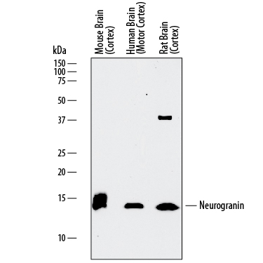
Detection of Human, Mouse, and Rat Neurogranin by Western Blot. Western blot shows lysates of mouse brain (cortex) tissue, human brain (motor cortex) tissue, and rat brain (cortex) tissue. PVDF membrane was probed with 0.5 µg/mL of Sheep Anti-Human/Mouse/Rat Neurogranin Antigen Affinity-purified Polyclonal Antibody (Catalog # AF7947) followed by HRP-conjugated Anti-Sheep IgG Secondary Antibody (HAF016). A specific band was detected for Neurogranin at approximately 14-16 kDa (as indicated). This experiment was conducted under reducing conditions and using Immunoblot Buffer Group 1.
 View Larger
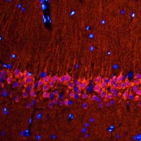
Neurogranin in Rat Brain. Neurogranin was detected in perfusion fixed frozen sections of rat brain (hippocampus) using Sheep Anti-Human/Mouse/Rat Neurogranin Antigen Affinity-purified Polyclonal Antibody (Catalog # AF7947) at 0.5 µg/mL overnight at 4 °C. Tissue was stained using the NorthernLights™ 557-conjugated Anti-Sheep IgG Secondary Antibody (red; NL010) and counterstained with DAPI (blue). Specific staining was localized to pyramidal neurons in the hippocampus. View our protocol for Fluorescent IHC Staining of Frozen Tissue Sections.
 View Larger
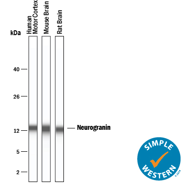
Detection of Human, Mouse, and Rat Neurogranin by Simple WesternTM. Simple Western lane view shows lysates of human brain (motor cortex), mouse brain, and rat brain, loaded at 0.2 mg/mL. A specific band was detected for Neurogranin at approximately 12 kDa (as indicated) using 20 µg/mL of Sheep Anti-Human/Mouse/Rat Neurogranin Antigen Affinity-purified Polyclonal Antibody (Catalog # AF7947) followed by 1:50 dilution of HRP-conjugated Anti-Sheep IgG Secondary Antibody (HAF016). This experiment was conducted under reducing conditions and using the 2-40 kDa separation system.
 View Larger
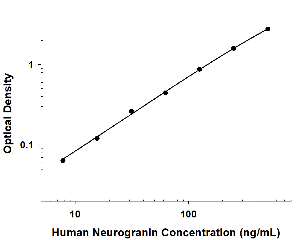
Human Neurogranin ELISA Standard Curve. Recombinant Human Neurogranin protein was serially diluted 2-fold and captured by Mouse Anti-Human Neurogranin Monoclonal Antibody (Catalog # MAB79473) coated on a Clear Polystyrene Microplate (Catalog # DY990). Sheep Anti-Human/Mouse/Rat Neurogranin Antigen Affinity-purified Polyclonal Antibody (Catalog # AF7947) was biotinylated and incubated with the protein captured on the plate. Detection of the standard curve was achieved by incubating Streptavidin-HRP (Catalog # DY998) followed by Substrate Solution (Catalog # DY999) and stopping the enzymatic reaction with Stop Solution (Catalog # DY994).
Reconstitution Calculator
Preparation and Storage
- 12 months from date of receipt, -20 to -70 °C as supplied.
- 1 month, 2 to 8 °C under sterile conditions after reconstitution.
- 6 months, -20 to -70 °C under sterile conditions after reconstitution.
Background: Neurogranin
NRGN (Neurogranin/Ng; also RC3, p17 and BICKS) is a member of the neurogranin family of proteins. Although its predicted MW is 7.5 kDa, it runs anomalously at 15-19 kDa in SDS-PAGE. This is apparently due to the adoption of a rigid alpha -helical structure in a negatively charged medium. NRGN has limited expression, being found principally in excitatory neurons of the telencephalon, Golgi and Purkinje cells of the cerebellum, and platelets plus B and T cells in non-nervous tissue. Intracellularly, NRGN is found associated with membranes of the ER, Golgi and mitochondria. This association is often in the form of aggregates (or granules), thus giving rise to its name ("neuro-granules"). NRGN is also found in the nucleus and associated with the postsynaptic spines of dendrites. The principal function of NRGN appears to be the binding, sequestration and concentration of CaM (calmodulin; a Ca-binding protein) in dendritic spines. Following NMDAR activation, Ca diffuses into the synaptic area, resulting in 1) the simply dissociation of CaM from NRGN, or 2) the phosphorylation of NRGN followed by its dissociation fro CaM. In either case, the freed CaM is now available to activate multiple downstream signaling pathways, some involved in LTP (or memory). Human NRGN is 78 amino acids (aa) in length. It contains one IQ domain (aa 26-47) that binds CaM, and a collagen-like region at the C-terminus (aa 48-78). Regulatory phosphorylation occurs on Ser36, and the N-terminal Met is acetylated. Full-length human NRGN shares 96% aa sequence identity with both mouse and rat NRGN.
Product Datasheets
Citation for Human/Mouse/Rat Neurogranin Antibody
R&D Systems personnel manually curate a database that contains references using R&D Systems products. The data collected includes not only links to publications in PubMed, but also provides information about sample types, species, and experimental conditions.
1 Citation: Showing 1 - 1
-
Regulation of prefrontal patterning and connectivity by retinoic acid
Authors: M Shibata, K Pattabiram, B Lorente-Ga, D Andrijevic, SK Kim, N Kaur, SK Muchnik, X Xing, G Santpere, AMM Sousa, N Sestan
Nature, 2021-10-01;0(0):.
Species: Human, Mouse
Sample Types: Whole Tissue
Applications: IHC
FAQs
No product specific FAQs exist for this product, however you may
View all Antibody FAQsReviews for Human/Mouse/Rat Neurogranin Antibody
There are currently no reviews for this product. Be the first to review Human/Mouse/Rat Neurogranin Antibody and earn rewards!
Have you used Human/Mouse/Rat Neurogranin Antibody?
Submit a review and receive an Amazon gift card.
$25/€18/£15/$25CAN/¥75 Yuan/¥2500 Yen for a review with an image
$10/€7/£6/$10 CAD/¥70 Yuan/¥1110 Yen for a review without an image

