Human/Mouse/Rat Ninjurin-2 Antibody Summary
Met1-Thr65
Accession # Q9NZG7
Applications
Please Note: Optimal dilutions should be determined by each laboratory for each application. General Protocols are available in the Technical Information section on our website.
Scientific Data
 View Larger
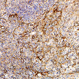
Detection of Ninjurin‑2 in Human Tonsil Ninjurin‑2 was detected in immersion fixed paraffin-embedded sections of Human Tonsil using Sheep Anti-Human/Mouse/Rat Ninjurin‑2 Antigen Affinity-purified Polyclonal Antibody (Catalog # AF5056) at 3 µg/mL for 1 hour at room temperature followed by incubation with the Anti-Sheep IgG VisUCyte™ HRP Polymer Antibody (Catalog # VC006). Before incubation with the primary antibody, tissue was subjected to heat-induced epitope retrieval using VisUCyte Antigen Retrieval Reagent-Basic (Catalog # VCTS021). Tissue was stained using DAB (brown) and counterstained with hematoxylin (blue). Specific staining was localized to cell surface on lymphocytes. View our protocol for IHC Staining with VisUCyte HRP Polymer Detection Reagents.
 View Larger
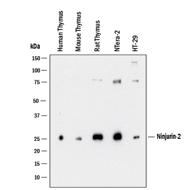
Detection of Human/Mouse/Rat Ninjurin‑2 by Western Blot. Western blot shows lysates of human thymus, mouse thymus, rat thymus, NTera‑2 human testicular embryonic carcinoma cells and HT‑29 human colon adenocarcinoma cells. PVDF membrane was probed with 1 µg/mL of Sheep Anti-Human/Mouse/Rat Ninjurin‑2 Antigen Affinity-purified Polyclonal Antibody (Catalog # AF5056) followed by HRP-conjugated Anti-Sheep IgG Secondary Antibody (Catalog # HAF016). A specific band was detected for Ninjurin‑2 at approximately ~26kDa kDa (as indicated). This experiment was conducted under reducing conditions and using Western Blot Buffer Group 1.
Reconstitution Calculator
Preparation and Storage
- 12 months from date of receipt, -20 to -70 °C as supplied.
- 1 month, 2 to 8 °C under sterile conditions after reconstitution.
- 6 months, -20 to -70 °C under sterile conditions after reconstitution.
Background: Ninjurin-2
Ninjurin-2 (nerve injury-induced protein 2) is a 20‑22 kDa member of the Ninjurin family of transmembrane (TM) proteins. It is expressed by multiple cell types, including Schwann cells, myenteric plexus and sensory neurons, and lymphocytes and participates in intercellular homophilic binding. Human Ninjurin-2 is 142 amino acids (aa) in length. It has an unusual membrane orientation. There is a 65 aa N-terminal extracellular domain (ECD) (aa 1‑65) that contains one phoshorylation site at Ser3, followed by a TM segment, a cytoplasmic region, a second TM segment and a C-terminal ECD (aa 128‑142). One potential alternate start site exists 46 aa upstream of the standard form start site. Over aa 1‑65, human Ninjurin-2 is 71% aa identical to mouse Ninjurin-2.
Product Datasheets
FAQs
No product specific FAQs exist for this product, however you may
View all Antibody FAQsReviews for Human/Mouse/Rat Ninjurin-2 Antibody
There are currently no reviews for this product. Be the first to review Human/Mouse/Rat Ninjurin-2 Antibody and earn rewards!
Have you used Human/Mouse/Rat Ninjurin-2 Antibody?
Submit a review and receive an Amazon gift card.
$25/€18/£15/$25CAN/¥75 Yuan/¥2500 Yen for a review with an image
$10/€7/£6/$10 CAD/¥70 Yuan/¥1110 Yen for a review without an image

