Human/Mouse/Rat nNOS Antibody Summary
Ser218-Ser1434
Accession # P29475
Applications
Please Note: Optimal dilutions should be determined by each laboratory for each application. General Protocols are available in the Technical Information section on our website.
Scientific Data
 View Larger
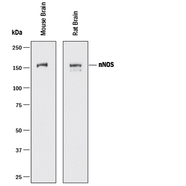
Detection of Mouse and Rat nNOS by Western Blot. Western blot shows lysates of mouse brain tissue and rat brain tissue. PVDF membrane was probed with 1 µg/mL of Goat Anti-Human/Mouse/Rat nNOS Antigen Affinity-purified Polyclonal Antibody (Catalog # AF2416) followed by HRP-conjugated Anti-Goat IgG Secondary Antibody (Catalog # HAF017). A specific band was detected for nNOS at approximately 160 kDa (as indicated). This experiment was conducted under reducing conditions and using Immunoblot Buffer Group 1.
 View Larger
nNOS in Human Brain. nNOS was detected in immersion fixed paraffin-embedded sections of human brain (cortex) using 1.7 µg/mL Goat Anti-Human/Mouse/Rat nNOS Antigen Affinity-purified Polyclonal Antibody (Catalog # AF2416) overnight at 4 °C. Tissue was stained with the Anti-Goat HRP-DAB Cell & Tissue Staining Kit (brown; Catalog # CTS008) and counterstained with hematoxylin (blue). Specific labeling was localized to the cytoplasm of astrocytes in the cortex. View our protocol for Chromogenic IHC Staining of Paraffin-embedded Tissue Sections.
 View Larger
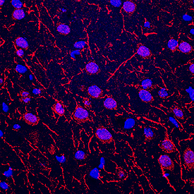
nNOS in Rat Brain. nNOS was detected in immersion fixed frozen sections of rat brain (cortex) using Goat Anti-Human/Mouse/Rat nNOS Antigen Affinity-purified Polyclonal Antibody (Catalog # AF2416) at 5 µg/mL overnight at 4 °C. Tissue was stained using the NorthernLights™ 557-conjugated Anti-Goat IgG Secondary Antibody (red; Catalog # NL001) and counterstained with DAPI (blue). Specific staining was localized to neurons and neuronal processes. View our protocol for Fluorescent IHC Staining of Frozen Tissue Sections.
 View Larger
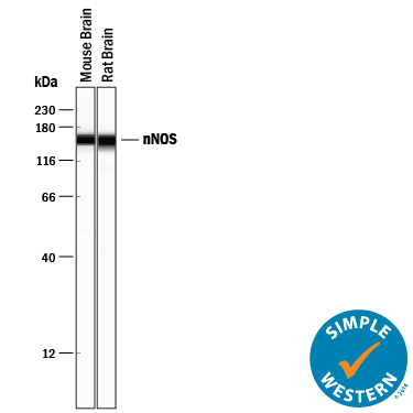
Detection of Mouse and Rat nNOS by Simple WesternTM. Simple Western lane view shows lysates of mouse brain tissue and rat brain tissue, loaded at 0.2 mg/mL. A specific band was detected for nNOS at approximately 160 kDa (as indicated) using 10 µg/mL of Goat Anti-Human/Mouse/Rat nNOS Antigen Affinity-purified Polyclonal Antibody (Catalog # AF2416) followed by 1:50 dilution of HRP-conjugated Anti-Goat IgG Secondary Antibody (Catalog # HAF109). This experiment was conducted under reducing conditions and using the 12-230 kDa separation system.
Reconstitution Calculator
Preparation and Storage
- 12 months from date of receipt, -20 to -70 °C as supplied.
- 1 month, 2 to 8 °C under sterile conditions after reconstitution.
- 6 months, -20 to -70 °C under sterile conditions after reconstitution.
Background: nNOS
nNOS is one of three NOS enzymes that catalyze the oxidation of L-argine to L-citruline and nitric oxide. nNOS exists as homodimers containing a cytochrome P450‑like prosthetic heme group in the N-terminal half. It also has a tightly bound FAD and FMN group in the C-terminal half. At least 4 isoforms of human nNOS are known. Human nNOS shares about 55% amino acid sequence identity with eNOS and iNOS. It also shares 96% sequence identity with mouse or rat nNOS.
Product Datasheets
FAQs
No product specific FAQs exist for this product, however you may
View all Antibody FAQsReviews for Human/Mouse/Rat nNOS Antibody
There are currently no reviews for this product. Be the first to review Human/Mouse/Rat nNOS Antibody and earn rewards!
Have you used Human/Mouse/Rat nNOS Antibody?
Submit a review and receive an Amazon gift card.
$25/€18/£15/$25CAN/¥75 Yuan/¥2500 Yen for a review with an image
$10/€7/£6/$10 CAD/¥70 Yuan/¥1110 Yen for a review without an image

