Human/Mouse/Rat Rap1A/B Antibody Summary
Met1-Leu184
Accession # P62834
Applications
Please Note: Optimal dilutions should be determined by each laboratory for each application. General Protocols are available in the Technical Information section on our website.
Scientific Data
 View Larger
Detection of Human Rap1A/B by Western Blot. Western blot shows recombinant human Rap1A, Rap1B, Rap2A, and Rap2B (1 ng/lane). PVDF membrane was probed with 1 µg/mL Human/Mouse/Rat Rap1A/B Antigen Affinity-purified Polyclonal Antibody (Catalog # AF3767) followed by HRP-conjugated Anti-Goat IgG Secondary Antibody (HAF017). This experiment was conducted under reducing conditions and using Immunoblot Buffer Group 1.
 View Larger
Detection of Human/Mouse/Rat Rap1A/B by Western Blot. Western blot shows lysates of K562 human chronic myelogenous leukemia cell line, MDA-MB-468 human breast cancer cell line, PT18 mouse mast/basophil cell line, and Rat-2 rat embryonic fibroblast cell line. PVDF membrane was probed with 1 µg/mL Human/Mouse/Rat Rap1A/B Antigen Affinity-purified Polyclonal Antibody (Catalog # AF3767) followed by HRP-conjugated Anti-Goat IgG Secondary Antibody (HAF017). A specific band for Rap1A/B was detected at approximately 22 kDa (as indicated). This experiment was conducted under reducing conditions and using Immunoblot Buffer Group 1.
 View Larger
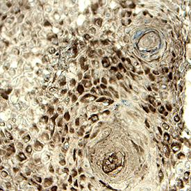
Detection of Rap1A/B in Human Squamous. Rap1A/B was detected in immersion fixed paraffin-embedded sections of Human Squamous using Goat Anti-Human/Mouse/Rat Rap1A/B Antigen Affinity-purified Polyclonal Antibody (Catalog # AF3767) at 15 µg/mL for 1 hour at room temperature followed by incubation with HRP-DAB. Before incubation with the primary antibody, tissue was subjected to heat-induced epitope retrieval using VisUCyte Antigen Retrieval Reagent-Basic (Catalog # VCTS021). Tissue was stained using DAB (brown) and counterstained with hematoxylin (blue). Specific staining was localized to cytoplasm of cancer cells. View our protocol for IHC Staining with VisUCyte HRP Polymer Detection Reagents.
Reconstitution Calculator
Preparation and Storage
- 12 months from date of receipt, -20 to -70 °C as supplied.
- 1 month, 2 to 8 °C under sterile conditions after reconstitution.
- 6 months, -20 to -70 °C under sterile conditions after reconstitution.
Background: Rap1A/B
The Ras-related proteins Rap1A and Rap1B share 95% identity and together with Rap2A and Rap2B constitute a grouping within the Ras superfamily of small GTPases. Like other GTPases, Rap proteins transduce signals by cycling between an active GTP-bound and an inactive GDP-bound state. Rap1 counteracts the mitogenic function of Ras, possibly through competitive interactions with Ras GAPs and Raf, and is implicated in the regulation of integrins.
Product Datasheets
Citation for Human/Mouse/Rat Rap1A/B Antibody
R&D Systems personnel manually curate a database that contains references using R&D Systems products. The data collected includes not only links to publications in PubMed, but also provides information about sample types, species, and experimental conditions.
1 Citation: Showing 1 - 1
-
Epstein-Barr virus subverts mevalonate and fatty acid pathways to promote infected B-cell proliferation and survival
Authors: LW Wang, Z Wang, I Ersing, L Nobre, R Guo, S Jiang, S Trudeau, B Zhao, MP Weekes, BE Gewurz
PLoS Pathog., 2019-09-13;15(9):e1008030.
Species: Human
Sample Types: Cell Lysates
Applications: Western Blot
FAQs
No product specific FAQs exist for this product, however you may
View all Antibody FAQsReviews for Human/Mouse/Rat Rap1A/B Antibody
There are currently no reviews for this product. Be the first to review Human/Mouse/Rat Rap1A/B Antibody and earn rewards!
Have you used Human/Mouse/Rat Rap1A/B Antibody?
Submit a review and receive an Amazon gift card.
$25/€18/£15/$25CAN/¥75 Yuan/¥2500 Yen for a review with an image
$10/€7/£6/$10 CAD/¥70 Yuan/¥1110 Yen for a review without an image

