Human/Mouse/Rat Syntaxin 7 Antibody Summary
Asn21-Glu187
Accession # O15400
Applications
Please Note: Optimal dilutions should be determined by each laboratory for each application. General Protocols are available in the Technical Information section on our website.
Scientific Data
 View Larger
Detection of Human Syntaxin 7 by Western Blot. Western blot shows recombinant human Syntaxin 12, 16, 1A, 1B2, 5, 6, 7, 8, and 1B1 (5 ng/lane). PVDF membrane was probed with 1 µg/mL Sheep Anti-Human/Mouse/Rat Syntaxin 7 Antigen Affinity-purified Polyclonal Antibody (Catalog # AF5478) followed by HRP-conjugated Anti-Sheep IgG Secondary Antibody (HAF016). A specific band for Syntaxin 7 was detected at approximately 29 kDa (as indicated). This experiment was conducted under reducing conditions and using Immunoblot Buffer Group 1.
 View Larger
Detection of Human, Mouse, and Rat Syntaxin 7 by Western Blot. Western blot shows lysates of BJAB human Burkitt's lymphoma cell line, MCF-7 human breast cancer cell line, C2C12 mouse myoblast cell line, BaF3 mouse pro-B cell line, L1.2 mouse pro-B cell line, and Rat-2 rat embryonic fibroblast cell line. PVDF membrane was probed with 1 µg/mL Sheep Anti-Human/Mouse/Rat Syntaxin 7 Antigen Affinity-purified Polyclonal Antibody (Catalog # AF5478) followed by HRP-conjugated Anti-Sheep IgG Secondary Antibody (HAF016). A specific band for Syntaxin 7 was detected at approximately 39 kDa (as indicated). This experiment was conducted under reducing conditions and using Immunoblot Buffer Group 1.
.") View Larger
View Larger
Syntaxin 7 in HeLa Human Cell Line. Syntaxin 7 was detected in immersion fixed HeLa human cervical epithelial carcinoma cell line using Sheep Anti-Human/Mouse/Rat Syntaxin 7 Antigen Affinity-purified Polyclonal Antibody (Catalog # AF5478) at 15 µg/mL for 3 hours at room temperature. Cells were stained using the NorthernLights™ 557-conjugated Anti-Sheep IgG Secondary Antibody (red; NL010) and counterstained with DAPI (blue). Specific staining was localized to lysosomes. View our protocol for Fluorescent ICC Staining of Cells on Coverslips.
 View Larger
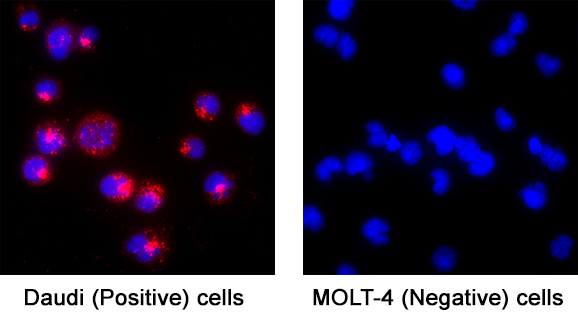
Detection of Syntaxin 7 in Daudi Human Burkitt's Lymphoma Cell Line (Positive) and MOLT‑4 Human Acute Lymphoblastic Leukemia Cell Line (Negative) Cells. Syntaxin 7 was detected in immersion fixed Daudi Human Burkitt's Lymphoma Cell Line (Positive) and MOLT‑4 Human Acute Lymphoblastic Leukemia Cell Line (Negative) Cells using Sheep Anti-Human/Mouse/Rat Syntaxin 7 Antigen Affinity-purified Polyclonal Antibody (Catalog # AF5478) at 5 µg/mL for 3 hours at room temperature. Cells were stained using the NorthernLights™ 557-conjugated Anti-Goat IgG Secondary Antibody (red; Catalog # NL001) and counterstained with DAPI (blue). Specific staining was localized to cytoplasm. View our protocol for Fluorescent ICC Staining of Non-adherent Cells.
Reconstitution Calculator
Preparation and Storage
- 12 months from date of receipt, -20 to -70 °C as supplied.
- 1 month, 2 to 8 °C under sterile conditions after reconstitution.
- 6 months, -20 to -70 °C under sterile conditions after reconstitution.
Background: Syntaxin 7
Syntaxin 7 (STX7) is a widely expressed protein embedded in endosomal and lysosomal membranes, and serves as a component of the SNARE complex. STX7 is involved in endocytic trafficking from early endosomes to late endosomes and lysosomes. This is in contrast to STX8, which is involved in clathrin-independent vesicular transport. Human STX7 is a type IV single-pass transmembrane protein (very short exoplasmic C-terminus) that is 261 amino acids (aa) in length. It contains a coiled-coil region (aa 47‑69), a t-SNARE domain (aa 165‑227) that is likely involved in protein-protein interactions, and a short, two amino acid, C-terminal luminal sequence. Over aa 21‑187, human STX7 shares 97% aa identity with mouse STX7.
Product Datasheets
Citation for Human/Mouse/Rat Syntaxin 7 Antibody
R&D Systems personnel manually curate a database that contains references using R&D Systems products. The data collected includes not only links to publications in PubMed, but also provides information about sample types, species, and experimental conditions.
1 Citation: Showing 1 - 1
-
Munc13-4 functions as a Ca2+ sensor for homotypic secretory granule fusion to generate endosomal exocytic vacuoles.
Authors: Sang Su Woo, Declan J James, Thomas F J Martin
Molecular Biology of the Cell, 2017-01-18;0(0):1939-4586.
Species: Rat
Sample Types: Cell Lysates
Applications: Western Blot
FAQs
No product specific FAQs exist for this product, however you may
View all Antibody FAQsReviews for Human/Mouse/Rat Syntaxin 7 Antibody
Average Rating: 4 (Based on 2 Reviews)
Have you used Human/Mouse/Rat Syntaxin 7 Antibody?
Submit a review and receive an Amazon gift card.
$25/€18/£15/$25CAN/¥75 Yuan/¥2500 Yen for a review with an image
$10/€7/£6/$10 CAD/¥70 Yuan/¥1110 Yen for a review without an image
Filter by:


