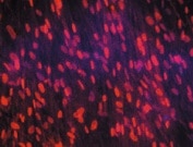
Human/Mouse/Rat YAP1 Antibody Summary
Applications
Please Note: Optimal dilutions should be determined by each laboratory for each application. General Protocols are available in the Technical Information section on our website.
Scientific Data
 View Larger
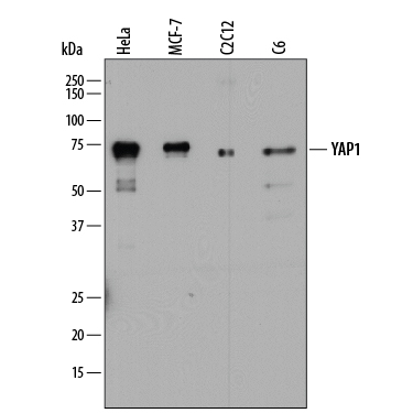
Detection of Human, Mouse, and Rat YAP1 by Western Blot. Western blot shows lysates of HeLa human cervical epithelial carcinoma cell line, MCF-7 human breast cancer cell line, C2C12 mouse myoblast cell line, and C6 rat glioma cell line. PVDF membrane was probed with 2 µg/mL of Mouse Anti-Human YAP1 Monoclonal Antibody (Catalog # MAB8094) followed by HRP-conjugated Anti-Mouse IgG Secondary Antibody (Catalog # HAF018). A specific band was detected for YAP1 at approximately 70-75 kDa (as indicated). This experiment was conducted under reducing conditions and using Immunoblot Buffer Group 1.
 View Larger
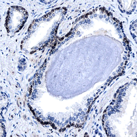
YAP1 in Human Prostate. YAP1 was detected in formalin fixed paraffin-embedded sections of human prostate using Mouse Anti-Human YAP1 Monoclonal Antibody (Catalog # MAB8094) at 15 µg/mL overnight at 4 °C. Before incubation with the primary antibody, tissue was subjected to heat-induced epitope retrieval using Antigen Retrieval Reagent-Basic (Catalog # CTS013). Tissue was stained using the Anti-Mouse HRP-DAB Cell & Tissue Staining Kit (brown; Catalog # CTS002) and counterstained with hematoxylin (blue). Specific staining was localized to the nuclei of smooth muscle cells. View our protocol for Chromogenic IHC Staining of Paraffin-embedded Tissue Sections.
 View Larger
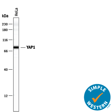
Detection of Human YAP1 by Simple WesternTM. Simple Western lane view shows lysates of HeLa human cervical epithelial carcinoma cell line, loaded at 0.5 mg/mL. A specific band was detected for YAP1 at approximately 79 kDa (as indicated) using 20 µg/mL of Mouse Anti-Human/Mouse/Rat YAP1 Monoclonal Antibody (Catalog # MAB8094). This experiment was conducted under reducing conditions and using the 12-230 kDa separation system.
 View Larger
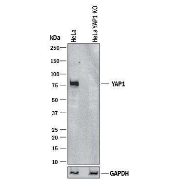
Western Blot Shows Human YAP1 Specificity by Using Knockout Cell Line. Western blot shows lysates of HeLa human cervical epithelial carcinoma parental cell line and YAP1 knockout HeLa cell line (KO). PVDF membrane was probed with 2 µg/mL of Mouse Anti-Human/Mouse/Rat YAP1 Monoclonal Antibody (Catalog # MAB8094) followed by HRP-conjugated Anti-Mouse IgG Secondary Antibody (Catalog # HAF018). A specific band was detected for YAP1 at approximately 75 kDa (as indicated) in the parental HeLa cell line, but is not detectable in knockout HeLa cell line. GAPDH (Catalog # MAB5718) is shown as a loading control. This experiment was conducted under reducing conditions and using Immunoblot Buffer Group 1.
Reconstitution Calculator
Preparation and Storage
- 12 months from date of receipt, -20 to -70 degreesC as supplied. 1 month, 2 to 8 degreesC under sterile conditions after reconstitution. 6 months, -20 to -70 degreesC under sterile conditions after reconstitution.
Background: YAP1
YAP1 (Yes-associated protein 1), also called YAP65 or Yorkie Homolog, is a widely expressed 65 kDa cytoplasmic and nuclear transcriptional regulator protein in the Hippo pathway that regulates organ size. The 504 amino acid (aa) human YAP1 contains two WW (tryptophan-containing protein binding) domains (aa 171-263), a transactivation domain (aa 219-504) and 19 sites that may be phosphorylated by kinases such as LATS1 and 2, MAPK8 and 9, CK1, or ABL1. Isoforms of 488, 454 and 326 aa lack aa 328-343, 230-267 (plus a 3 aa substitution for aa 329-343), or 1-178, respectively. YAP1 binds the Yes tyrosine kinase SH3 domain and many other proteins, notably ErbB4. YAP1 expression is increased in some liver and prostate cancers. Within aa 347-504, human YAP1 shares 94% and 93% aa sequence identity with mouse and rat YAP1, respectively.
Product Datasheets
Citations for Human/Mouse/Rat YAP1 Antibody
R&D Systems personnel manually curate a database that contains references using R&D Systems products. The data collected includes not only links to publications in PubMed, but also provides information about sample types, species, and experimental conditions.
3
Citations: Showing 1 - 3
Filter your results:
Filter by:
-
Immunohistochemistry of YAP and dNp63 and survival analysis of patients bearing precancerous lesion and oral squamous cell carcinoma
Authors: S Ono, K Nakano, K Takabatake, H Kawai, H Nagatsuka
Int J Med Sci, 2019-05-28;16(5):766-773.
Species: Human
Sample Types: Whole Tissue
Applications: IHC -
Antibody-drug conjugate T-DM1 treatment for HER2+ breast cancer induces ROR1 and confers resistance through activation of Hippo transcriptional coactivator YAP1
Authors: SS Islam, M Uddin, ASM Noman, H Akter, NJ Dity, M Basiruzzma, F Uddin, J Ahsan, S Annoor, AA Alaiya, M Al-Alwan, H Yeger, WA Farhat
EBioMedicine, 2019-05-10;0(0):.
Species: Human
Sample Types: Cell Lysates, Whole Cells
Applications: ICC, Western Blot -
A connexin43/YAP axis regulates astroglial-mesenchymal transition in hemoglobin induced astrocyte activation
Authors: Y Yang, J Ren, Y Sun, Y Xue, Z Zhang, A Gong, B Wang, Z Zhong, Z Cui, Z Xi, GY Yang, Q Sun, L Bian
Cell Death Differ., 2018-06-07;0(0):.
Species: Rat
Sample Types: Protein
Applications: Immunoprecipitation
FAQs
No product specific FAQs exist for this product, however you may
View all Antibody FAQsReviews for Human/Mouse/Rat YAP1 Antibody
Average Rating: 5 (Based on 1 Review)
Have you used Human/Mouse/Rat YAP1 Antibody?
Submit a review and receive an Amazon gift card.
$25/€18/£15/$25CAN/¥75 Yuan/¥2500 Yen for a review with an image
$10/€7/£6/$10 CAD/¥70 Yuan/¥1110 Yen for a review without an image
Filter by:
