Human MYH7 Antibody Summary
Accession # P12883
Applications
Please Note: Optimal dilutions should be determined by each laboratory for each application. General Protocols are available in the Technical Information section on our website.
Scientific Data
 View Larger
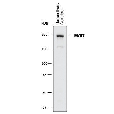
Detection of MYH7 by Western Blot. Western blot shows lysates of human heart (ventricle) tissue. PVDF membrane was probed with 0.5 µg/mL of Rabbit Anti-Human MYH7 Monoclonal Antibody (Catalog # MAB90961) followed by HRP-conjugated Anti-Rabbit IgG Secondary Antibody (Catalog # HAF008). A specific band was detected for MYH7 at approximately 230 kDa (as indicated). This experiment was conducted under reducing conditions and using Immunoblot Buffer Group 1.
 View Larger
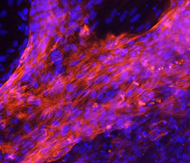
MYH7 in Human Cardiomyocytes. MYH7 was detected in immersion fixed BG01V human embryonic stem cells differentiated into cardiomyocytes using Rabbit Anti-Human MYH7 Monoclonal Antibody (Catalog # MAB90961) at 10 µg/mL for 3 hours at room temperature. Cells were stained using the NorthernLights™ 557-conjugated Anti-Rabbit IgG Secondary Antibody (red; Catalog # NL004) and counterstained with DAPI (blue). Specific staining was localized to cytoplasm. View our protocol for Fluorescent ICC Staining of Stem Cells on Coverslips.
Reconstitution Calculator
Preparation and Storage
- 12 months from date of receipt, -20 to -70 °C as supplied.
- 1 month, 2 to 8 °C under sterile conditions after reconstitution.
- 6 months, -20 to -70 °C under sterile conditions after reconstitution.
Background: MYH7
MYH7 encodes the beta myosin heavy chain (MHC-beta ) which is a component of cardiac muscle myosin mainly expressed in the ventricle of fetal heart and represents the minority myosin in the adult heart. This is the 'slow form' of cardiac myosin as opposed to the 'fast form' (MYH6, aka MHC-alpha ) expressed more predominantely in the atria of the fetal heart and is the predominant myosin in the adult heart. The two isoforms of cardiac MHC alpha and beta display 93% homology but have significantly different enzymatic properties, with alpha having 150-300% the contractile velocity and 60-70% actin attachment time as that of beta. Several mutations in MYH7 have been associated with inherited cardiomyopathies paraspinal and proximal muscle atrophy. MYH7 is a 223 kDa protein composed of 1935 amino acids.
Product Datasheets
Product Specific Notices
Contains <0.1% Sodium Azide, which is not hazardous at this concentration according to GHS classifications. Refer to SDS for additional information and handling instructions.Citations for Human MYH7 Antibody
R&D Systems personnel manually curate a database that contains references using R&D Systems products. The data collected includes not only links to publications in PubMed, but also provides information about sample types, species, and experimental conditions.
2
Citations: Showing 1 - 2
Filter your results:
Filter by:
-
Histone acetyltransferase Kat2a regulates ferroptosis via enhancing Tfrc and Hmox1 expression in diabetic cardiomyopathy
Authors: Zhen, J;Sheng, X;Chen, T;Yu, H;
Cell death & disease
Species: Mouse
Sample Types: Tissue Homogenates, Cell Lysates
Applications: Western Blot -
An essential role for Wnt/?-catenin signaling in mediating hypertensive heart disease
Authors: Y Zhao, C Wang, C Wang, X Hong, J Miao, Y Liao, L Zhou, Y Liu
Sci Rep, 2018-06-12;8(1):8996.
Species: Rat
Sample Types: Tissue Homogenates
Applications: Western Blot
FAQs
No product specific FAQs exist for this product, however you may
View all Antibody FAQsReviews for Human MYH7 Antibody
Average Rating: 4 (Based on 1 Review)
Have you used Human MYH7 Antibody?
Submit a review and receive an Amazon gift card.
$25/€18/£15/$25CAN/¥75 Yuan/¥2500 Yen for a review with an image
$10/€7/£6/$10 CAD/¥70 Yuan/¥1110 Yen for a review without an image
Filter by:
the antibody was used for immunofluorescence staining on day 15 human iPSC-derived cardiomyocytes (the day of initiation of differentiation from iPSCs is counted as day 0).


