Human NFkB2 Antibody Summary
Met1-Asn447
Accession # Q00653
Applications
Please Note: Optimal dilutions should be determined by each laboratory for each application. General Protocols are available in the Technical Information section on our website.
Scientific Data
 View Larger
Detection of Human NF kappa B2 by Western Blot. Western blot shows lysates of Daudi human Burkitt's lymphoma cell line untreated (-) or treated (+) with 100 ng/mL Recombinant Human CD40 Ligand/TNFSF5 aa 108-261 (Catalog # 6245-CL) for 4 hours. Gels were loaded with 20 µg of cytoplasmic (Cyto) and 10 µg of nuclear extracts (Nuc). For additional reference, lysates of Raji human Burkitt's lymphoma cell line were included. PVDF membrane was probed with 0.5 µg/mL Mouse Anti-Human NF kappa B2 Monoclonal Antibody (Catalog # MAB28881) followed by HRP-conjugated Anti-Mouse IgG Secondary Antibody (Catalog # HAF007). Specific bands for NF kappa B2 were detected at approximately 52 kDa and 100 kDa (as indicated). This experiment was conducted under reducing conditions and using Immunoblot Buffer Group 1.
 View Larger
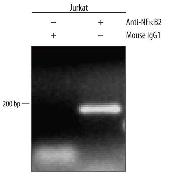
Detection of NF kappa B2-regulated Genes by Chromatin Immunoprecipitation. Jurkat human acute T cell leukemia cell line treated with 50 ng/mL PMA and 200 ng/mL calcium ionomycin overnight was fixed using formaldehyde, resuspended in lysis buffer, and sonicated to shear chromatin. NF kappa B2/DNA complexes were immunoprecipitated using 5 µg Mouse Anti-Human NF kappa B2 Monoclonal Antibody (Catalog # MAB28881) or control antibody (Catalog # MAB002) for 15 minutes in an ultrasonic bath, followed by Biotinylated Anti-Mouse IgG Secondary Antibody (Catalog # BAF007). Immunocomplexes were captured using 50 µL of MagCellect Streptavidin Ferrofluid (Catalog # MAG999) and DNA was purified using chelating resin solution. Thec-mycpromoter was detected by standard PCR.
 View Larger
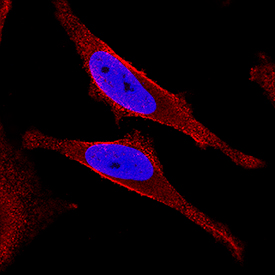
NF kappa B2 in HeLa Human Cell Line. NF kappa B2 was detected in immersion fixed HeLa human cervical epithelial carcinoma cell line using Mouse Anti-Human NF kappa B2 Monoclonal Antibody (Catalog # MAB28881) at 3 µg/mL for 3 hours at room temperature. Cells were stained using the NorthernLights™ 557-conjugated Anti-Mouse IgG Secondary Antibody (red; Catalog # NL007) and counterstained with DAPI (blue). Specific staining was localized to cytoplasm and nuclei. View our protocol for Fluorescent ICC Staining of Cells on Coverslips.
Reconstitution Calculator
Preparation and Storage
- 12 months from date of receipt, -20 to -70 °C as supplied.
- 1 month, 2 to 8 °C under sterile conditions after reconstitution.
- 6 months, -20 to -70 °C under sterile conditions after reconstitution.
Background: NFkB2
Nuclear Factor kappa B2 (NF kappa B2 or NF kappa B p52) is a member of the NF kappa B/Rel family of transcription factors. NF kappa B2 dimerizes with other members of the NF kappa B/Rel family to regulate expression of genes that participate in immune, apoptotic, and oncogenic processes.
Product Datasheets
FAQs
No product specific FAQs exist for this product, however you may
View all Antibody FAQsReviews for Human NFkB2 Antibody
Average Rating: 4.2 (Based on 5 Reviews)
Have you used Human NFkB2 Antibody?
Submit a review and receive an Amazon gift card.
$25/€18/£15/$25CAN/¥75 Yuan/¥2500 Yen for a review with an image
$10/€7/£6/$10 CAD/¥70 Yuan/¥1110 Yen for a review without an image
Filter by:
Antibody was printed on custom arrays and incubated with fluorescently labeled human EDTA plasma
Down-regulation of NFKB-2 p100/p52 in Human melanoma A375 cell line upon treatment with ethanol. Dilution: 1:1,000 in PBS with 5% BSA. Secondary Ab: anti-Mouse IgG 1:5,000.


