Human NKp46/NCR1 Antibody Summary
Gln22-Asn254
Accession # AAH64806
Applications
Please Note: Optimal dilutions should be determined by each laboratory for each application. General Protocols are available in the Technical Information section on our website.
Scientific Data
 View Larger
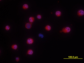
NKp46/NCR1 in NK‑92 human cell line NKp46/NCR1 was detected in immersion fixed NK-92 human natural killer lymphoma cell line using Goat Anti-Human NKp46/NCR1 Antigen Affinity-purified Polyclonal Antibody (Catalog # AF1850) at 10 µg/mL for 3 hours at room temperature. Cells were stained using the NorthernLights™ 557-conjugated Anti-Goat IgG Secondary Antibody (red; Catalog # NL001) and counterstained with DAPI (blue). View our protocol for Fluorescent ICC Staining of Non-adherent Cells.
 View Larger
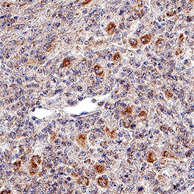
NKp46/NCR1 in Human Lymph Node. NKp46/NCR1 was detected in immersion fixed paraffin-embedded sections of human lymph node using Goat Anti-Human NKp46/NCR1 Antigen Affinity-purified Polyclonal Antibody (Catalog # AF1850) at 3 µg/mL for 1 hour at room temperature followed by incubation with the Anti-Goat IgG VisUCyte™ HRP Polymer Antibody (Catalog # VC004). Tissue was stained using DAB (brown) and counterstained with hematoxylin (blue). Specific staining was localized to cytoplasm. View our protocol for IHC Staining with VisUCyte HRP Polymer Detection Reagents.
 View Larger
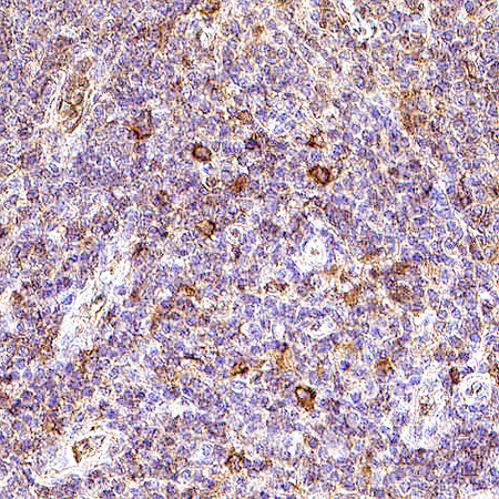
NKp46/NCR1 in Human Lymph Node. NKp46/NCR1 was detected in immersion fixed paraffin-embedded sections of human lymph node using Goat Anti-Human NKp46/NCR1 Antigen Affinity-purified Polyclonal Antibody (Catalog # AF1850) at 8 µg/mL for 1 hour at room temperature followed by incubation with the Anti-Goat IgG VisUCyte™ HRP Polymer Antibody (VC004). Before incubation with the primary antibody, tissue was subjected to heat-induced epitope retrieval using Antigen Retrieval Reagent-Basic (CTS013). Tissue was stained using DAB (brown) and counterstained with hematoxylin (blue). Specific staining was localized to cytoplasm in lymphocytes.
Reconstitution Calculator
Preparation and Storage
- 12 months from date of receipt, -20 to -70 °C as supplied.
- 1 month, 2 to 8 °C under sterile conditions after reconstitution.
- 6 months, -20 to -70 °C under sterile conditions after reconstitution.
Background: NKp46/NCR1
NKp46, along with NKp30 and NKp44, are activating receptors that have been collectively termed the natural cytotoxicity receptors (NCR) (1). These receptors lack significant sequence homology to one another. They are expressed almost exclusively by NK cells and play a major role in triggering some of the key lytic activities of NK cells. The CD56dimCD16+ subpopulation that makes up the majority of NK cells in the peripheral blood and spleen expresses NKp46 in both resting and activated states (2). The main NK cell population of the lymph node (CD56brightCD16-) expresses low levels of NKp46 in resting cells, but expression is up-regulated by IL-2. NKp46 is a type I transmembrane protein with two extracellular Ig-like domains followed by a short stalk region, a transmembrane domain containing a positively charged amino acid residue, and a short cytoplasmic tail. Through its positive charge in the transmembrane domain, NKp46 associates with the
ITAM-bearing signal adapter proteins, CD3 zeta and Fc epsilon R1 gamma, which are able to form disulfide-linked homodimers and heterodimers (3, 8). Studies with neutralizing antibodies indicate that the three NCRs are primarily responsible for triggering the NK-mediated lysis of many human tumor cell lines. Blocking any of the NCRs individually resulted in partial inhibition of tumor cell lysis, but nearly complete inhibition of lysis was observed if all three receptors were blocked simultaneously (4). NKp46 has also been implicated in recognition of virus-infected cells through its capacity to bind to viral hemagglutinins (5-7). Human NKp46 shares 58% and 59% amino acid sequence identity with the mouse and rat proteins, respectively.
- Moretta, L. and A. Moretta (2004) EMBO J. 23:255.
- Ferlazzo, G. et al. (2004) J. Immunol. 172:1455.
- Augugliaro, R. et al. (2003) Eur. J. Immunol. 33:1235.
- Pende, D. et al. (1999) J. Exp. Med. 190:1505.
- Arnon, T. et al. (2004) Blood 103:664.
- Arnon, T. et al. (2001) Eur. J. Immunol. 31:2680.
- Mandelboim, O. et al. (2001) Nature 409:1055.
- Moretta, A. et al. (2001) Annu. Rev. Immunol. 19:197.
Product Datasheets
Citations for Human NKp46/NCR1 Antibody
R&D Systems personnel manually curate a database that contains references using R&D Systems products. The data collected includes not only links to publications in PubMed, but also provides information about sample types, species, and experimental conditions.
6
Citations: Showing 1 - 6
Filter your results:
Filter by:
-
Dipeptidyl Peptidase 4 Inhibitors Reduce Hepatocellular Carcinoma by Activating Lymphocyte Chemotaxis in Mice
Authors: S Nishina, A Yamauchi, T Kawaguchi, K Kaku, M Goto, K Sasaki, Y Hara, Y Tomiyama, F Kuribayash, T Torimura, K Hino
Cell Mol Gastroenterol Hepatol, 2018-09-11;7(1):115-134.
Species: Human
Sample Types: Whole Tissue
Applications: IHC-P -
Evidence of innate lymphoid cell redundancy in humans
Authors: F Vély, V Barlogis, B Vallentin, B Neven, C Piperoglou, M Ebbo, T Perchet, M Petit, N Yessaad, F Touzot, J Bruneau, N Mahlaoui, N Zucchini, C Farnarier, G Michel, D Moshous, S Blanche, A Dujardin, H Spits, JH Distler, A Ramming, C Picard, R Golub, A Fischer, E Vivier
Nat. Immunol, 2016-09-12;17(11):1291-1299.
Species: Human
Sample Types: Whole Tissue
Applications: IHC -
Fc receptor-mediated phagocytosis in tissues as a potent mechanism for preventive and therapeutic HIV vaccine strategies
Mucosal Immunol, 2016-02-17;9(6):1584-1595.
Species: Human
Sample Types: Whole Tissue
Applications: IHC-P -
In situ characterization of intrahepatic non-parenchymal cells in PSC reveals phenotypic patterns associated with disease severity.
Authors: Berglin L, Bergquist A, Johansson H, Glaumann H, Jorns C, Lunemann S, Wedemeyer H, Ellis E, Bjorkstrom N
PLoS ONE, 2014-08-20;9(8):e105375.
Species: Human
Sample Types: Whole Tissue
Applications: IHC-Fr -
Up-regulation of a death receptor renders antiviral T cells susceptible to NK cell-mediated deletion.
Authors: Peppa, Dimitra, Gill, Upkar S, Reynolds, Gary, Easom, Nicholas, Pallett, Laura J, Schurich, Anna, Micco, Lorenzo, Nebbia, Gaia, Singh, Harsimra, Adams, David H, Kennedy, Patrick, Maini, Mala K
J Exp Med, 2012-12-17;210(1):99-114.
Species: Human
Sample Types: Whole Tissue
Applications: IHC-P -
Innate immunity in multiple sclerosis white matter lesions: expression of natural cytotoxicity triggering receptor 1 (NCR1).
Authors: Durrenberger PF, Ettorre A, Kamel F
J Neuroinflammation, 2012-01-02;9(0):1.
Species: Human
Sample Types: Whole Cells, Whole Tissue
Applications: ICC, IHC-Fr
FAQs
No product specific FAQs exist for this product, however you may
View all Antibody FAQsReviews for Human NKp46/NCR1 Antibody
There are currently no reviews for this product. Be the first to review Human NKp46/NCR1 Antibody and earn rewards!
Have you used Human NKp46/NCR1 Antibody?
Submit a review and receive an Amazon gift card.
$25/€18/£15/$25CAN/¥75 Yuan/¥2500 Yen for a review with an image
$10/€7/£6/$10 CAD/¥70 Yuan/¥1110 Yen for a review without an image


