Human NT-3 Antibody Summary
Tyr139-Thr257
Accession # P20783
Applications
Please Note: Optimal dilutions should be determined by each laboratory for each application. General Protocols are available in the Technical Information section on our website.
Scientific Data
 View Larger
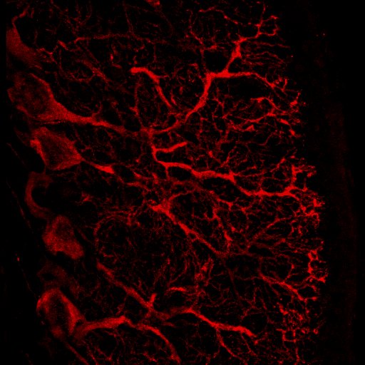
NT‑3 in Rat Brain. NT‑3 was detected in perfusion fixed frozen sections of rat brain (Purkinje cells in the cerebellum) using 10 µg/mL Human NT‑3 Antigen Affinity-purified Polyclonal Antibody (Catalog # AF‑267‑NA) overnight at 4 °C. Tissue was stained with the Anti-Goat HRP-AEC Cell & Tissue Staining Kit (red; Catalog # CTS009) and counterstained with hematoxylin (blue). View our protocol for Chromogenic IHC Staining of Paraffin-embedded Tissue Sections.
 View Larger
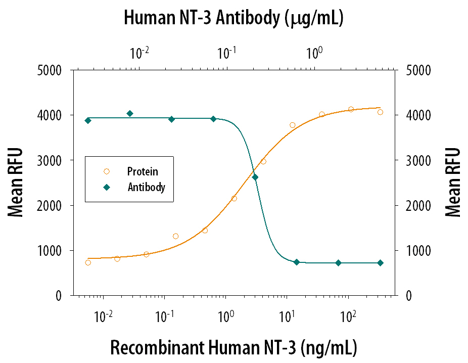
Cell Proliferation Induced by NT‑3 and Neutralization by Human NT‑3 Antibody. Recombinant Human NT-3 (Catalog # 267-N3) stimulates proliferation in BaF-TrKB-BD mouse pro-B cell line transfected with TrkB in a dose-dependent manner (orange line). Proliferation elicited by Recombinant Human NT-3 (20 ng/mL) is neutralized (green line) by increasing concentrations of Human NT-3 Antigen Affinity-purified Polyclonal Antibody (Catalog # AF-267-NA). The ND50 is typically 0.1-0.4 µg/mL.
Reconstitution Calculator
Preparation and Storage
- 12 months from date of receipt, -20 to -70 °C as supplied.
- 1 month, 2 to 8 °C under sterile conditions after reconstitution.
- 6 months, -20 to -70 °C under sterile conditions after reconstitution.
Background: NT-3
Neurotrophin-3 (NT-3) is a member of the NGF family of neurotrophic factors (also named neurotrophins) that are required for the differentiation and survival of specific neuronal subpopulations in both the central as well as the peripheral nervous systems. The neurotrophin family is comprised of at least four proteins including NGF, BDNF, NT-3, and NT-4/5. These secreted cytokines are synthesized as prepropeptides that are proteolytically processed to generate the mature proteins. All neurotrophins have six conserved cysteine residues that are involved in the formation of three disulfide bonds and all share approximately 55% sequence identity at the amino acid level. Similarly to NGF, bioactive NT-3 is predicted to be a non-covalently linked homodimer.
The NT-3 cDNA encodes a 257 amino acid residue precursor protein with a signal peptide and a proprotein that are cleaved to yield the 119 amino acid residue mature NT-3. The amino acid sequence of mature NT-3 is identical in human, mouse and rat. NT-3 transcripts have been detected in the cerebellum, hippocampus, placenta, heart, skin, and skeletal muscle. NT-3 primarily activates the TrkC tyrosine kinase receptor. In addition, NT-3 can activate Trk and TrkB kinase receptors in certain cell systems. NT-3 can also bind with low affinity to the low affinity NGF receptor.
- Eide, F.F. et al. (1993) Exp. Neurol. 121:200.
- Snider, W.D. (1994) Cell 77:627.
- Barbacid, M. (1994) J. Neurobiol. 25:1386.
Product Datasheets
Citation for Human NT-3 Antibody
R&D Systems personnel manually curate a database that contains references using R&D Systems products. The data collected includes not only links to publications in PubMed, but also provides information about sample types, species, and experimental conditions.
1 Citation: Showing 1 - 1
-
Electrical stimulation of sympathetic neurons induces autocrine/paracrine effects of NGF mediated by TrkA.
Authors: Saygili E, Schauerte P, Kuppers F, Heck L, Weis J, Weber C, Schwinger RH, Hoffmann R, Schroder JW, Marx N, Rana OR
J. Mol. Cell. Cardiol., 2010-02-02;49(1):79-87.
Species: Rat
Sample Types: Whole Cells
Applications: Neutralization
FAQs
No product specific FAQs exist for this product, however you may
View all Antibody FAQsReviews for Human NT-3 Antibody
Average Rating: 4.5 (Based on 2 Reviews)
Have you used Human NT-3 Antibody?
Submit a review and receive an Amazon gift card.
$25/€18/£15/$25CAN/¥75 Yuan/¥2500 Yen for a review with an image
$10/€7/£6/$10 CAD/¥70 Yuan/¥1110 Yen for a review without an image
Filter by:


