Human PD-1 Antibody Summary
Leu25-Gln167
Accession # Q8IX89
Applications
Please Note: Optimal dilutions should be determined by each laboratory for each application. General Protocols are available in the Technical Information section on our website.
Scientific Data
 View Larger
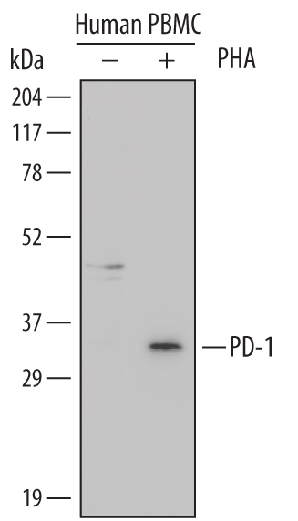
Detection of Human PD‑1 by Western Blot. Western blot shows lysates of human peripheral blood mononuclear cells (PBMC) untreated (-) or treated (+) with 1 µg/mL PHA for 5 days. PVDF Membrane was probed with 1 µg/mL of Human PD-1 Monoclonal Antibody (Catalog # MAB1086) followed by HRP-conjugated Anti-Mouse IgG Secondary Antibody (Catalog # HAF007). A specific band was detected for PD-1 at approximately 33 kDa (as indicated). This experiment was conducted under reducing conditions and using Immunoblot Buffer Group 1.
Reconstitution Calculator
Preparation and Storage
- 12 months from date of receipt, -20 to -70 °C as supplied.
- 1 month, 2 to 8 °C under sterile conditions after reconstitution.
- 6 months, -20 to -70 °C under sterile conditions after reconstitution.
Background: PD-1
Programmed Death-1 (PD-1) is a type I transmembrane protein belonging to the CD28/CTLA-4 family of immunoreceptors that mediate signals for regulating immune responses (1). Members of the CD28/CTLA-4 family have been shown to either promote T cell activation (CD28 and ICOS) or downregulate T cell activation (CTLA-4 and PD-1) (2). PD-1 is expressed on activated T cells, B cells, myeloid cells, and on a subset of thymocytes. In vitro, ligation of PD-1 inhibits TCR-mediated T cell proliferation and production of IL-1, IL-4, IL-10, and IFN-gamma. In addition, PD-1 ligation also inhibits BCR mediated signaling. PD-1 deficient mice have a defect in peripheral tolerance and spontaneously develop autoimmune diseases (2, 3). Two B7 family proteins, PD-L1 (also called B7-H1) and PD-L2 (also known as B7-DC), have been identified as PD-1 ligands. Unlike other B7 family proteins, both PD‑L1 and PD‑L2 are expressed in a wide variety of normal tissues including heart, placenta, and activated spleens (4). The wide expression of PD-L1 and PD-L2 and the inhibitor effects on PD-1 ligation indicate that PD-1 might be involved in the regulation of peripheral tolerance and may help prevent autoimmune diseases (2). The human PD-1 gene encodes a 288 amino acid (aa) protein with a putative 20 aa signal peptide, a 148 aa extracellular region with one immunoglobulin-like V‑type domain, a 24 aa transmembrane domain, and a 95 aa cytoplasmic region. The cytoplasmic tail contains two tyrosine residues that form the immunoreceptor tyrosine-based inhibitory motif (ITIM) and immunoreceptor tyrosine-based switch motif (ITSM) that are important in mediating PD-1 signaling. Mouse and human PD-1 share approximately 60% aa sequence identity (4).
- Ishida, Y. et al. (1992) EMBO J. 11:3887.
- Nishimura, H. and T. Honjo (2001) Trends in Immunol. 22:265.
- Latchman, Y. et al. (2001) Nature Immun. 2:261.
- Carreno, B.M. and M. Collins (2002) Annu. Rev. Immunol. 20:29.
Product Datasheets
Citations for Human PD-1 Antibody
R&D Systems personnel manually curate a database that contains references using R&D Systems products. The data collected includes not only links to publications in PubMed, but also provides information about sample types, species, and experimental conditions.
5
Citations: Showing 1 - 5
Filter your results:
Filter by:
-
Evaluation of the immune checkpoint factors in idiopathic membranous nephropathy
Authors: R Motavalli, M Hosseini, MS Soltani-Za, A Karimi, M Sadeghi, S Dolati, M Yousefi, J Etemadi
Molecular and cellular probes, 2023-04-21;69(0):101914.
Species: Human
Sample Types: Cell Lysates
Applications: Western Blot -
Lymph node migratory dendritic cells modulate HIV-1 transcription through PD-1 engagement
Authors: R Banga, C Rebecchini, FA Procopio, A Noto, O Munoz, K Ioannidou, C Fenwick, K Ohmiti, M Cavassini, JM Corpataux, L de Leval, G Pantaleo, M Perreau
PLoS Pathog., 2019-07-22;15(7):e1007918.
Species: Human
Sample Types: Whole Tissue
Applications: IHC-P -
Soluble co-signaling molecules predict long-term graft outcome in kidney-transplanted patients.
Authors: Melendreras S, Martinez-Camblor P, Menendez A, Bravo-Mendoza C, Gonzalez-Vidal A, Coto E, Diaz-Corte C, Ruiz-Ortega M, Lopez-Larrea C, Suarez-Alvarez B
PLoS ONE, 2014-12-05;9(12):e113396.
Species: Human
Sample Types: Serum
Applications: ELISA Development -
Splenic proliferative lymphoid nodules distinct from germinal centers are sites of autoantigen stimulation in immune thrombocytopenia.
Authors: Daridon C, Loddenkemper C, Spieckermann S, Kuhl A, Salama A, Burmester G, Lipsky P, Dorner T
Blood, 2012-09-06;120(25):5021-31.
Species: Human
Sample Types: Whole Tissue
Applications: IHC-P -
Programmed Death-1: from gene to protein in autoimmune human myasthenia gravis.
Authors: Sakthivel P, Ramanujam R, Wang XB, Pirskanen R, Lefvert AK
J. Neuroimmunol., 2007-11-26;193(1):149-55.
Species: Human
Sample Types: Serum
Applications: ELISA Development
FAQs
No product specific FAQs exist for this product, however you may
View all Antibody FAQsReviews for Human PD-1 Antibody
Average Rating: 4 (Based on 1 Review)
Have you used Human PD-1 Antibody?
Submit a review and receive an Amazon gift card.
$25/€18/£15/$25CAN/¥75 Yuan/¥2500 Yen for a review with an image
$10/€7/£6/$10 CAD/¥70 Yuan/¥1110 Yen for a review without an image
Filter by:
293T cells were infected with letivirus carrying human PD-1 or control for 72h. Total cell lysates were collected and subject to Western Blot. HeLa protein sample was used as a PD-1 positive control. Membrane was probed with 1 µg/mL of Human PD‑1 Monoclonal Antibody (Catalog # MAB1086). PD‑1 band was detected at approximately 33 kDa


