Human PD-L1/B7-H1 Antibody Summary
Phe19-Thr239
Accession # Q9NZQ7
Applications
This antibody functions as an ELISA detection antibody when paired with Mouse Anti-Human PD-L1/B7-H1 Monoclonal Antibody (Catalog # MAB1561R).
This product is intended for assay development on various assay platforms requiring antibody pairs. We recommend the Human PD-L1/B7-H1 DuoSet ELISA Kit (Catalog # DY156) for convenient development of a sandwich ELISA or the Human/Cynomolgus Monkey PD-L1/B7-H1 Quantikine ELISA Kit (Catalog # DB7H10) for a complete optimized ELISA.
Please Note: Optimal dilutions should be determined by each laboratory for each application. General Protocols are available in the Technical Information section on our website.
Scientific Data
 View Larger
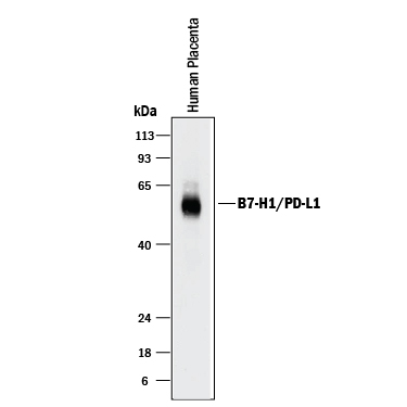
Detection of Human PD-L1/B7-H1 by Western Blot. Western blot shows lysates of human placenta tissue. PVDF membrane was probed with 2 µg/mL of Goat Anti-Human PD-L1/B7-H1 Antigen Affinity-purified Polyclonal Antibody (Catalog # AF156) followed by HRP-conjugated Anti-Goat IgG Secondary Antibody (Catalog # HAF017). A specific band was detected for PD-L1/B7-H1 at approximately 50-55 kDa (as indicated). This experiment was conducted under reducing conditions and using Immunoblot Buffer Group 1.
 View Larger
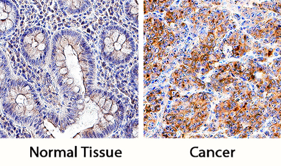
PD-L1/B7-H1 in Human Colon and Colon Cancer Tissue. PD-L1/B7-H1 was detected in immersion fixed paraffin-embedded sections of normal human colon (left panel) and human colon cancer tissue (right panel) using Goat Anti-Human PD-L1/B7-H1 Antigen Affinity-purified Polyclonal Antibody (Catalog # AF156) at 5 µg/mL overnight at 4 °C. Tissue was stained using the Anti-Goat HRP-DAB Cell & Tissue Staining Kit (brown; Catalog # CTS008) and counterstained with hematoxylin (blue). Specific staining was localized to cell membranes and cytoplasm. View our protocol for Chromogenic IHC Staining of Paraffin-embedded Tissue Sections.
 View Larger
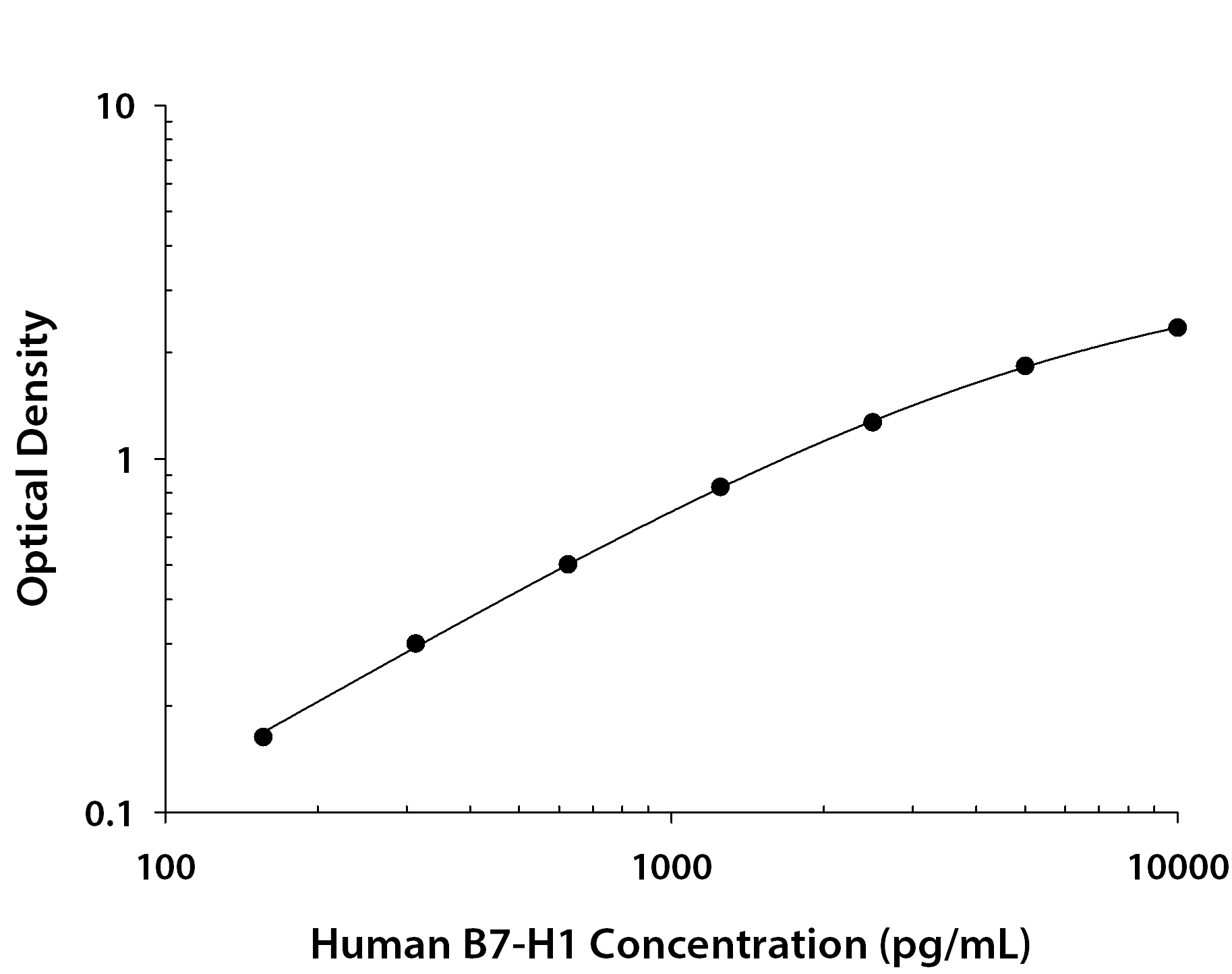
Human PD-L1/B7-H1 ELISA Standard Curve. Recombinant Human PD-L1/B7-H1 protein was serially diluted 2-fold and captured by Mouse Anti-Human PD-L1/B7-H1 Monoclonal Antibody (Catalog # MAB1561R) coated on a Clear Polystyrene Microplate (Catalog # DY990). Goat Anti-Human PD-L1/B7-H1 Antigen Affinity-purified Polyclonal Antibody (Catalog # AF156) was biotinylated and incubated with the protein captured on the plate. Detection of the standard curve was achieved by incubating Streptavidin-HRP (Catalog # DY998) followed by Substrate Solution (Catalog # DY999) and stopping the enzymatic reaction with Stop Solution (Catalog # DY994).
 View Larger
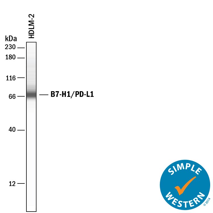
Detection of Human PD-L1/B7-H1 by Simple WesternTM. Simple Western lane view shows lysates of HDLM-2 human Hodgkin's lymphoma cell line, loaded at 0.2 mg/mL. A specific band was detected for PD-L1/B7-H1 at approximately 72 kDa (as indicated) using 50 µg/mL of Goat Anti-Human PD-L1/B7-H1 Antigen Affinity-purified Polyclonal Antibody (Catalog # AF156) followed by 1:50 dilution of HRP-conjugated Anti-Goat IgG Secondary Antibody (Catalog # HAF109). This experiment was conducted under reducing conditions and using the 12-230 kDa separation system.
Reconstitution Calculator
Preparation and Storage
- 12 months from date of receipt, -20 to -70 °C as supplied.
- 1 month, 2 to 8 °C under sterile conditions after reconstitution.
- 6 months, -20 to -70 °C under sterile conditions after reconstitution.
Background: PD-L1/B7-H1
Human B7 homolog 1 (B7-H1), also called programmed cell death 1 ligand 1 (PDCD1L1) and programmed death ligand 1 (PDL1), is a member of the growing B7 family of immune proteins that provide signals for both stimulating and inhibiting T cell activation. Other family members include B7-1, B7-2, B7-H2, PDL2 and B7-H3. B7 proteins are members of the immunoglobulin (Ig) superfamily, their extracellular domains contain 2 Ig-like domains and all members have short cytoplasmic domains. Among the family members, they share about 20-25% amino acid identity. Human and mouse B7-H1 share approximately 70% amino acid sequence identity. B7-H1 has been identified as one of two ligands for programmed death-1 (PD-1), a member of the CD28 family of immunoreceptors. The B7-H1 gene encodes a 290 amino acid (aa) type I membrane precursor protein with a putative 18 aa signal peptide, a 221 aa extracellular domain, a 21 aa transmembrane region, and a 31 aa cytoplasmic domain. Human B7-H1 is constitutively expressed in several organs such as heart, skeletal muscle, placenta and lung, and in lower amounts in thymus, spleen, kidney and liver. B7-H1 expression is upregulated in a small fraction of activated T and B cells and a much larger fraction of activated monocytes. B7-H1 expression is also induced in dendritic cells and keratinocytes after IFN-gamma stimulation. Interaction of B7-H1 with PD-1 results in inhibition of TCR-mediated proliferation and cytokine production. The B7-H1:PD-1 pathway is involved in the negative regulation of some immune responses and may play an important role in the regulation of peripheral tolerance.
- Nishimura, H. and T. Honjo (2001) Trends in Immunology 22:265.
- Freeman, G.J. et al. (2000) J. Exp. Med. 192:1027.
- Latchman, Y. et al. (2001) Nat. Immunol. 2:261.
Product Datasheets
Citations for Human PD-L1/B7-H1 Antibody
R&D Systems personnel manually curate a database that contains references using R&D Systems products. The data collected includes not only links to publications in PubMed, but also provides information about sample types, species, and experimental conditions.
18
Citations: Showing 1 - 10
Filter your results:
Filter by:
-
Small Extracellular Vesicles Harboring PD-L1 in Obstructive Sleep Apnea
Authors: Recoquillon, S;Ali, S;Justeau, G;Riou, J;Martinez, MC;Andriantsitohaina, R;Gagnadoux, F;Trzepizur, W;
International journal of molecular sciences
Species: Human hepegivirus
Sample Types: Cell Lysates, Extracellular Vesicles
Applications: Western Blot -
Interference with pathways activated by topoisomerase inhibition alters the surface expression of PD-L1 and MHC I in colon cancer cells
Authors: M Hassan, V Trung, D Bedi, S Shaddox, D Gunturu, C Yates, P Datta, T Samuel
Oncology Letters, 2022-12-09;25(1):41.
Species: Human
Sample Types: Whole Cells
Applications: Flow Cytometry -
STAG2 regulates interferon signaling in melanoma via enhancer loop reprogramming
Authors: Z Chu, L Gu, Y Hu, X Zhang, M Li, J Chen, D Teng, M Huang, CH Shen, L Cai, T Yoshida, Y Qi, Z Niu, A Feng, S Geng, DT Frederick, E Specht, A Piris, RJ Sullivan, KT Flaherty, GM Boland, K Georgopoul, D Liu, Y Shi, B Zheng
Nature Communications, 2022-04-06;13(1):1859.
Species: Human
Sample Types: Cell Lysates
Applications: Western Blot -
Adipose-Tissue-Derived Mesenchymal Stem Cells Mediate PD-L1 Overexpression in the White Adipose Tissue of Obese Individuals, Resulting in T Cell Dysfunction
Authors: A Eljaafari, J Pestel, B Le Maguere, S Chanon, J Watson, M Robert, E Disse, H Vidal
Cells, 2021-10-03;10(10):.
Species: Human
Sample Types: Whole Cells
Applications: Neutralization -
Immune checkpoint molecules B7-H6 and PD-L1 co-pattern the tumor inflammatory microenvironment in human breast cancer
Authors: B Cherif, H Triki, S Charfi, L Bouzidi, WB Kridis, A Khanfir, K Chaabane, T Sellami-Bo, A Rebai
Scientific Reports, 2021-04-06;11(1):7550.
Species: Human
Sample Types: Whole Tissue
Applications: IHC -
Checkpoint inhibition through small molecule-induced internalization of programmed death-ligand 1
Authors: JJ Park, EP Thi, VH Carpio, Y Bi, AG Cole, BD Dorsey, K Fan, T Harasym, CL Iott, S Kadhim, JH Kim, ACH Lee, D Nguyen, BS Paratala, R Qiu, A White, D Lakshminar, C Leo, RK Suto, R Rijnbrand, S Tang, MJ Sofia, CB Moore
Nature Communications, 2021-02-22;12(1):1222.
Species: Mouse
Sample Types: Cell Lysates
Applications: Western Blot -
Human amnion-derived mesenchymal stem cells attenuate xenogeneic graft-versus-host disease by preventing T cell activation and proliferation
Authors: Y Tago, C Kobayashi, M Ogura, J Wada, S Yamaguchi, T Yamaguchi, M Hayashi, T Nakaishi, H Kubo, Y Ueda
Scientific Reports, 2021-01-28;11(1):2406.
Species: Human
Sample Types: Whole Cells
Applications: Neutralization -
The innate immune effector ISG12a promotes cancer immunity by suppressing the canonical Wnt/&beta-catenin signaling pathway
Authors: R Deng, C Zuo, Y Li, B Xue, Z Xun, Y Guo, X Wang, Y Xu, R Tian, S Chen, Q Liu, J Chen, J Wang, X Huang, H Li, M Guo, X Wang, M Yang, Z Wu, J Wang, J Ma, J Hu, G Li, S Tang, Z Tu, H Ji, H Zhu
Cell. Mol. Immunol., 2020-09-22;0(0):.
Species: Human
Sample Types: Whole Cells
Applications: Neutralization -
Mechanisms utilized by feline adipose-derived mesenchymal stem cells to inhibit T lymphocyte proliferation
Authors: N Taechangam, SS Iyer, NJ Walker, B Arzi, DL Borjesson
Stem Cell Res Ther, 2019-06-25;10(1):188.
Species: Feline
Sample Types: Whole Cells
Applications: Flow Cytometry -
Immunological Properties of Human Embryonic Stem Cell-Derived Retinal Pigment Epithelial Cells
Authors: M Idelson, R Alper, A Obolensky, N Yachimovic, J Rachmilewi, A Ejzenberg, E Beider, E Banin, B Reubinoff
Stem Cell Reports, 2018-08-16;0(0):.
Species: Human
Sample Types: Whole Cells
Applications: Neutralization -
Restoration of T Cell function in multi-drug resistant bacterial sepsis after interleukin-7, anti-PD-L1, and OX-40 administration
Authors: LK Thampy, KE Remy, AH Walton, Z Hong, K Liu, R Liu, V Yi, CD Burnham, RS Hotchkiss
PLoS ONE, 2018-06-26;13(6):e0199497.
Species: Human
Sample Types: Whole Cells
Applications: Inhibition -
Blocking the PD-1/PD-L1 pathway in glioma: a potential new treatment strategy
Authors: S Xue, M Hu, V Iyer, J Yu
J Hematol Oncol, 2017-04-07;10(1):81.
Species: Human
Sample Types: Tissue Homogenates
Applications: Western Blot -
Hepatitis C Virus Induces MDSCs-Like Monocytes through TLR2/PI3K/AKT/STAT3 Signaling
Authors: N Zhai, H Li, H Song, Y Yang, A Cui, T Li, J Niu, IN Crispe, L Su, Z Tu
PLoS ONE, 2017-01-23;12(1):e0170516.
Species: Human
Sample Types: Whole Cells
Applications: Bioassay -
Mesenchymal Stromal Cell Secretion of Programmed Death-1 Ligands Regulates T Cell Mediated Immunosuppression
Stem Cells, 2016-10-26;0(0):.
Species: Human
Sample Types: Whole Cells
Applications: Immunoprecipitation -
Soluble co-signaling molecules predict long-term graft outcome in kidney-transplanted patients.
Authors: Melendreras S, Martinez-Camblor P, Menendez A, Bravo-Mendoza C, Gonzalez-Vidal A, Coto E, Diaz-Corte C, Ruiz-Ortega M, Lopez-Larrea C, Suarez-Alvarez B
PLoS ONE, 2014-12-05;9(12):e113396.
Species: Human
Sample Types: Serum
Applications: ELISA Development -
Overexpression of B7-H1 correlates with malignant cell proliferation in pancreatic cancer.
Authors: Song, Xiao, Liu, Junwei, Lu, Yi, Jin, Hongchua, Huang, Dongshen
Oncol Rep, 2013-12-31;31(3):1191-8.
Species: Human
Sample Types: Protein
Applications: Western Blot -
12-O-tetradecanoyl phorbol 13-acetate induces the expression of B7-DC, -H1, -H2, and -H3 in K562 cells.
Authors: Jang BC, Park YK, Choi IH, Kim SP, Hwang JB, Baek WK, Suh MH, Mun KC, Suh SI
Int. J. Oncol., 2007-12-01;31(6):1439-47.
Species: Human
Sample Types: Whole Cells
Applications: Flow Cytometry -
Aberrant regulation of synovial T cell activation by soluble costimulatory molecules in rheumatoid arthritis.
Authors: Wan B, Nie H, Liu A, Feng G, He D, Xu R, Zhang Q, Dong C, Zhang JZ
J. Immunol., 2006-12-15;177(12):8844-50.
Species: Human
Sample Types: Serum
Applications: ELISA Development
FAQs
No product specific FAQs exist for this product, however you may
View all Antibody FAQsReviews for Human PD-L1/B7-H1 Antibody
Average Rating: 4.7 (Based on 3 Reviews)
Have you used Human PD-L1/B7-H1 Antibody?
Submit a review and receive an Amazon gift card.
$25/€18/£15/$25CAN/¥75 Yuan/¥2500 Yen for a review with an image
$10/€7/£6/$10 CAD/¥70 Yuan/¥1110 Yen for a review without an image
Filter by:


