Human Phospho-Akt1 (T308) Antibody Summary
Applications
Please Note: Optimal dilutions should be determined by each laboratory for each application. General Protocols are available in the Technical Information section on our website.
Scientific Data
 View Larger
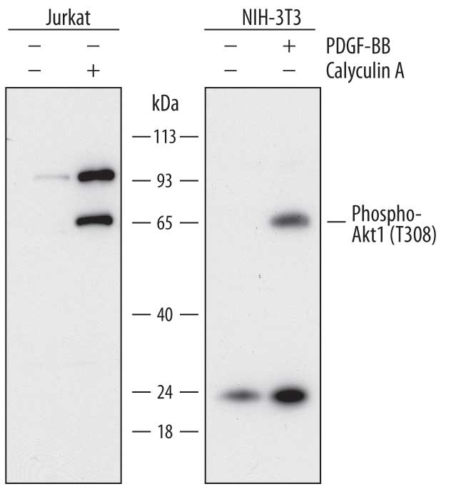
Detection of Human and Mouse Phospho-Akt1 (T308) by Western Blot. Western blot shows lysates of Jurkat human acute T cell leukemia cell line and NIH-3T3 mouse embryonic fibroblast cell line untreated (-) or treated (+) with 100 ng/mL Recombinant Human PDGF-BB (Catalog # 220-BB) for 20 minutes or 100nM Calyculin A for 30 minutes. PVDF membrane was probed with 1 µg/mL of Mouse Anti-Human Phospho-Akt1 (T308) Monoclonal Antibody (Catalog # MAB7419) followed by HRP-conjugated Anti-Mouse IgG Secondary Antibody (Catalog # HAF018). A specific band was detected for Phospho-Akt1 (T308) at approximately 65 kDa (as indicated). This experiment was conducted under reducing conditions and using Immunoblot Buffer Group 1.
 View Larger
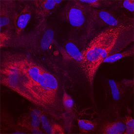
Phospho-Akt1 (T308) in CCD‑1070Sk Human Cell Line. Akt1 phosphorylated at T308 was detected in immersion fixed CCD-1070Sk human foreskin fibroblast cell line stimulated with Recombinant Human PDGF-BB (Catalog # 220-BB) using Mouse Anti-Human Phospho-Akt1 (T308) Mono-clonal Antibody (Catalog # MAB7419) at 25 µg/mL for 3 hours at room temperature. Cells were stained using the Northern-Lights™ 557-conjugated Anti-Mouse IgG Secondary Antibody (red; Catalog # NL007) and counterstained with DAPI (blue). Specific staining was localized to cytoplasm. View our protocol for Fluorescent ICC Staining of Cells on Coverslips.
Reconstitution Calculator
Preparation and Storage
- 12 months from date of receipt, -20 to -70 °C as supplied.
- 1 month, 2 to 8 °C under sterile conditions after reconstitution.
- 6 months, -20 to -70 °C under sterile conditions after reconstitution.
Background: Akt1
Akt, also known as protein kinase B (PKB), is a central kinase in such diverse cellular processes as glucose uptake, cell cycle progression, and apoptosis. Three highly homologous members define the Akt family: Akt1 (PKB alpha ), Akt2 (PKB beta ), and Akt3 (PKB gamma ). All three Akts contain an amino-terminal pleckstrin homology domain, a central kinase domain, and a carboxyl-terminal regulatory domain. Akt1 is the most widely expressed family member and is frequently activated in a number of carcinomas, including breast, prostate, lung, pancreatic, liver, ovarian, and colorectal cancer. Akt1 is activated in a multistep process that involves the sequential phosphorylation of Thr450 by JNK kinases, Thr308 by PDK1, and Ser473 by PDK2 or mTORC2. Activated Akt1 phosphorylates a wide variety of cytosolic, nuclear, and mitochondrial substrates. Human Akt1 shares 98% aa sequence identity with mouse and rat Akt1. MAB7419 also detects mouse Phospho-Akt1 (T308) in Western Blot.
Product Datasheets
Citations for Human Phospho-Akt1 (T308) Antibody
R&D Systems personnel manually curate a database that contains references using R&D Systems products. The data collected includes not only links to publications in PubMed, but also provides information about sample types, species, and experimental conditions.
3
Citations: Showing 1 - 3
Filter your results:
Filter by:
-
A Three-Dimensional Xeno-Free Culture Condition for Wharton's Jelly-Mesenchymal Stem Cells: The Pros and Cons
Authors: B Koh, N Sulaiman, MB Fauzi, JX Law, MH Ng, TL Yuan, AGN Azurah, MH Mohd Yunus, RBH Idrus, MD Yazid
International Journal of Molecular Sciences, 2023-02-13;24(4):.
Species: Human
Sample Types: Cell Lysates
Applications: Western Blot -
Distinct molecular pathways mediate Mycn and Myc-regulated miR-17-92 microRNA action in Feingold syndrome mouse models
Authors: F Mirzamoham, A Kozlova, G Papaioanno, E Paltrinier, UM Ayturk, T Kobayashi
Nat Commun, 2018-04-10;9(1):1352.
Species: Mouse
Sample Types: Cell Lysates
Applications: Western Blot -
ASPP2 Is a Novel Pan-Ras Nanocluster Scaffold
PLoS ONE, 2016-07-20;11(7):e0159677.
Species: Human
Sample Types: Cell Lysates
Applications: Western Blot
FAQs
No product specific FAQs exist for this product, however you may
View all Antibody FAQsReviews for Human Phospho-Akt1 (T308) Antibody
There are currently no reviews for this product. Be the first to review Human Phospho-Akt1 (T308) Antibody and earn rewards!
Have you used Human Phospho-Akt1 (T308) Antibody?
Submit a review and receive an Amazon gift card.
$25/€18/£15/$25CAN/¥75 Yuan/¥2500 Yen for a review with an image
$10/€7/£6/$10 CAD/¥70 Yuan/¥1110 Yen for a review without an image


