Human PRAS40 Antibody Summary
Applications
Please Note: Optimal dilutions should be determined by each laboratory for each application. General Protocols are available in the Technical Information section on our website.
Scientific Data
 View Larger
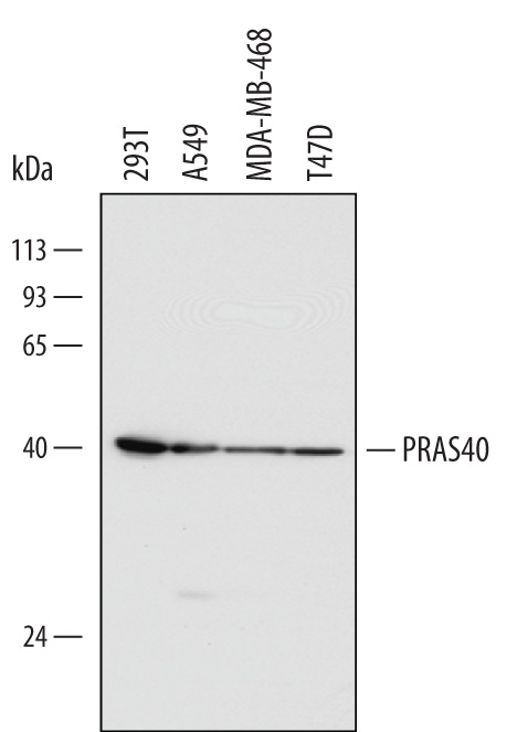
Detection of Human PRAS40 by Western Blot. Western blot shows lysates of 293T human embryonic kidney cell line, A549 human lung carcinoma cell line, MDA-MB-468 human breast cancer cell line, and T47D human breast cancer cell line. PVDF Membrane was probed with 1 µg/mL of Mouse Anti-Human PRAS40 Monoclonal Antibody (Catalog # MAB6408) followed by HRP-conjugated Anti-Mouse IgG Secondary Antibody (Catalog # HAF007). A specific band was detected for PRAS40 at approximately 40 kDa (as indicated). This experiment was conducted under reducing conditions and using Immunoblot Buffer Group 1.
 View Larger
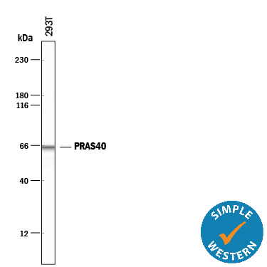
Detection of Human PRAS40 by Simple WesternTM. Simple Western lane view shows lysates of 293T human embryonic kidney cell line, loaded at 0.5 mg/mL. A specific band was detected for PRAS40 at approximately 65 kDa (as indicated) using 10 µg/mL of Mouse Anti-Human PRAS40 Monoclonal Antibody (Catalog # MAB6408). This experiment was conducted under reducing conditions and using the 12-230 kDa separation system.
 View Larger
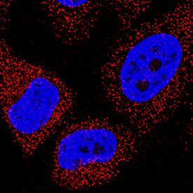
PRAS40 in HeLa Human Cell Line. PRAS40 was detected in immersion fixed HeLa human cervical epithelial carcinoma cell line using Mouse Anti-Human PRAS40 Monoclonal Antibody (Catalog # MAB6408) at 8 µg/mL for 3 hours at room temperature. Cells were stained using the NorthernLights™ 557-conjugated Anti-Mouse IgG Secondary Antibody (red; Catalog # NL007) and counterstained with DAPI (blue). Specific staining was localized to cytoplasm. View our protocol for Fluorescent ICC Staining of Cells on Coverslips.
Reconstitution Calculator
Preparation and Storage
- 12 months from date of receipt, -20 to -70 degreesC as supplied. 1 month, 2 to 8 degreesC under sterile conditions after reconstitution. 6 months, -20 to -70 degreesC under sterile conditions after reconstitution.
Background: PRAS40
PRAS40 (Proline-rich AKT1 substrate 1; also Akt1S1 and p39) is a 40-42 kDa cytoplasmic phosphoprotein that lacks generally recognized structural motifs. It is widely expressed and is considered to be key regulator of mTORC1 (mTOR plus raptor and G beta L), a complex through which Akt signals into the cell. Through phosphorylation, mTORC1 activity is upregulated by PRAS40. In particular, nonphosphorylated PRAS40 binds to and serves as a negative regulator of mTORC1 activity. Upon Insulin signaling, PRAS40 is phosphorylated on Thr246, Ser221 and Ser183. This causes it to bind 14-3-3 and results in its dissociation from mTORC1, freeing up mTOR to regulate (positively or negatively) protein synthesis. Human PRAS40 is 256 amino acids (aa) in length. It contains one extended Pro-rich region (aa 35-96) plus at least nine utilized Ser/Thr phosphorylation sites. There is one alternative start site at Met131. Over aa 119-256, human PRAS40 shares 93% aa identity with mouse PRAS40.
Product Datasheets
Citation for Human PRAS40 Antibody
R&D Systems personnel manually curate a database that contains references using R&D Systems products. The data collected includes not only links to publications in PubMed, but also provides information about sample types, species, and experimental conditions.
1 Citation: Showing 1 - 1
-
Soluble bone-derived osteopontin promotes migration and stem-like behavior of breast cancer cells
Authors: GM Pio, Y Xia, MM Piaseczny, JE Chu, AL Allan
PLoS ONE, 2017-05-12;12(5):e0177640.
Species: Human
Sample Types: Cell Lysates
Applications: Western Blot
FAQs
No product specific FAQs exist for this product, however you may
View all Antibody FAQsReviews for Human PRAS40 Antibody
There are currently no reviews for this product. Be the first to review Human PRAS40 Antibody and earn rewards!
Have you used Human PRAS40 Antibody?
Submit a review and receive an Amazon gift card.
$25/€18/£15/$25CAN/¥75 Yuan/¥2500 Yen for a review with an image
$10/€7/£6/$10 CAD/¥70 Yuan/¥1110 Yen for a review without an image


