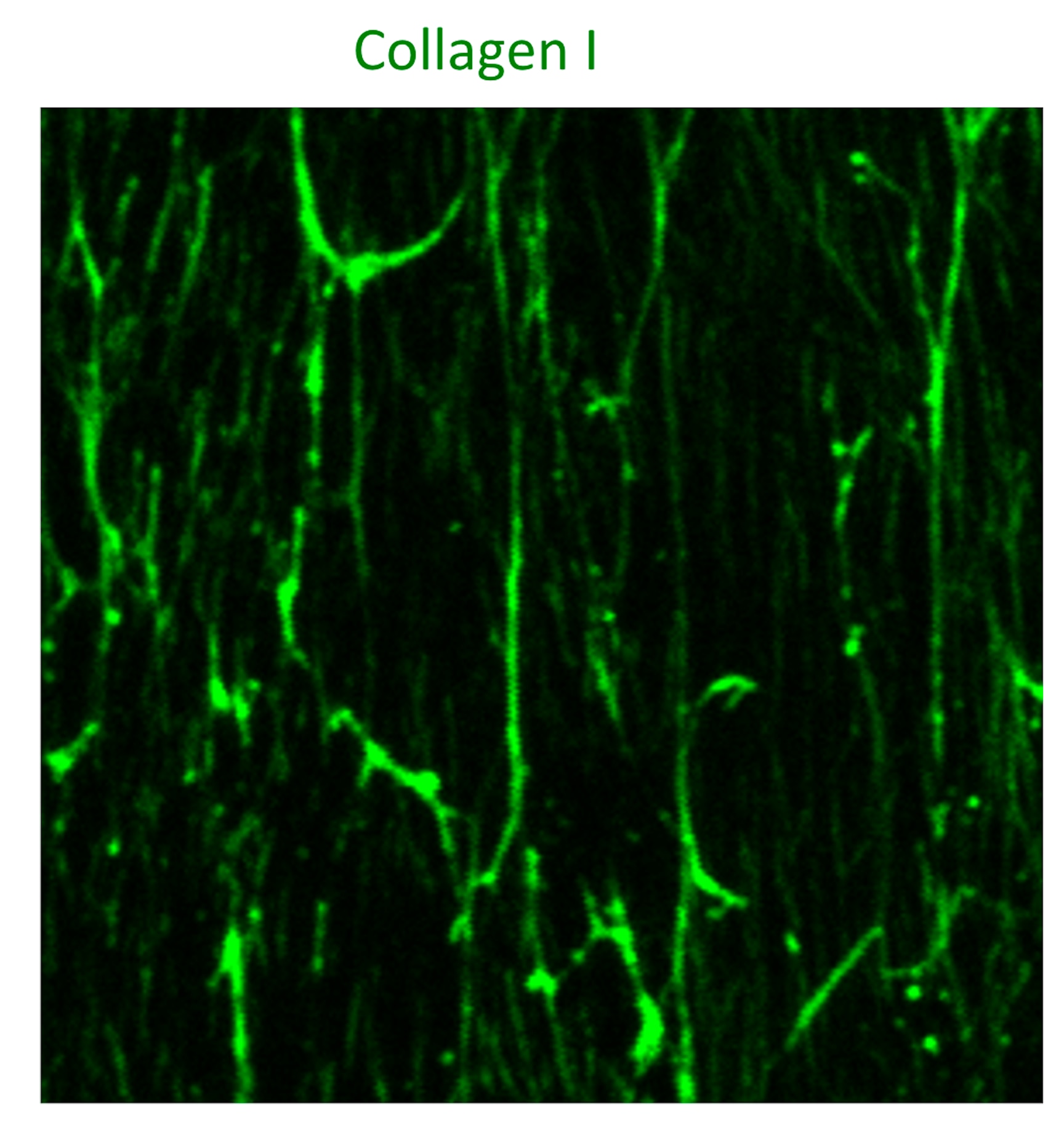
Human Pro Collagen I alpha 1 Antibody Summary
Gln23-Lys277, Gly1094-Leu1464
Accession # P02452
Applications
Please Note: Optimal dilutions should be determined by each laboratory for each application. General Protocols are available in the Technical Information section on our website.
Scientific Data
 View Larger
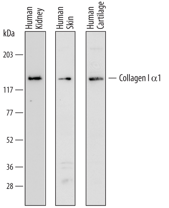
Detection of Human Pro-Collagen I alpha 1 by Western Blot. Western blot shows lysates of human kidney tissue, human skin tissue, and human cartilage tissue. PVDF membrane was probed with 1 µg/mL of Sheep Anti-Human Pro-Collagen I alpha 1 Antigen Affinity-purified Polyclonal Antibody (Catalog # AF6220) followed by HRP-conjugated Anti-Sheep IgG Secondary Antibody (Catalog # HAF016). A specific band was detected for Pro-Collagen I alpha 1 at approximately 140 kDa (as indicated). This experiment was conducted under reducing conditions and using Immunoblot Buffer Group 1.
 View Larger
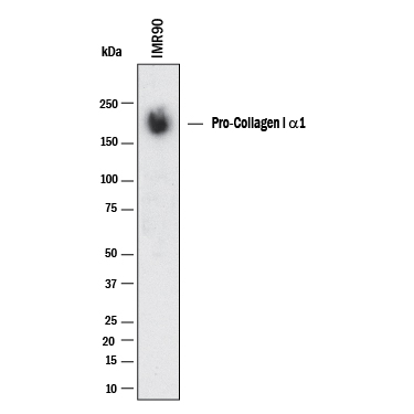
Detection of Human Collagen I by Western Blot. Western blot shows lysates of IMR‑90 human lung fibroblast cell line. PVDF membrane was probed with 1 µg/mL of Sheep Anti-Human Pro Collagen I alpha 1 Antigen Affinity-purified Polyclonal Antibody (Catalog # AF6220) followed by HRP-conjugated Anti-Sheep IgG Secondary Antibody (HAF016). A specific band was detected for Collagen I at approximately 170 kDa (as indicated). This experiment was conducted under reducing conditions and using Western Blot Buffer Group 1.
 View Larger
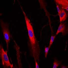
Pro-Collagen I alpha 1 in IMR‑90 Human Cell Line. Pro-Collagen I alpha 1 was detected in immersion fixed IMR-90 human lung fibroblast cell line using Sheep Anti-Human Pro-Collagen I alpha 1 Antigen Affinity-purified Polyclonal Antibody (Catalog # AF6220) at 10 µg/mL for 3 hours at room temperature. Cells were stained using the NorthernLights™ 557-conjugated Anti-Sheep IgG Secondary Antibody (red; Catalog # NL010) and counterstained with DAPI (blue). View our protocol for Fluorescent ICC Staining of Cells on Coverslips.
 View Larger
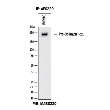
Detection of Human N-Pro Collagen I by Immunoprecipitation. Human N-Pro Collagen I was immunoprecipitated from 500 μg of IMR‑90 human lung fibroblast cell line lysates with 5 μg of Sheep Anti-Human N-Pro Collagen I Antigen Affinity-purified Polyclonal Antibody (Catalog # AF6220). The N-Pro Collagen I-antibody complexes were absorbed using Protein A or Protein G. Immunoprecipitated human N-Pro Collagen I was detected by Western blot using 1 μg/mL of Mouse Anti-Human N-Pro Collagen I Monoclonal Antibody (MAB6220) under reducing conditions and using Western Blot Buffer Group 1.
 View Larger
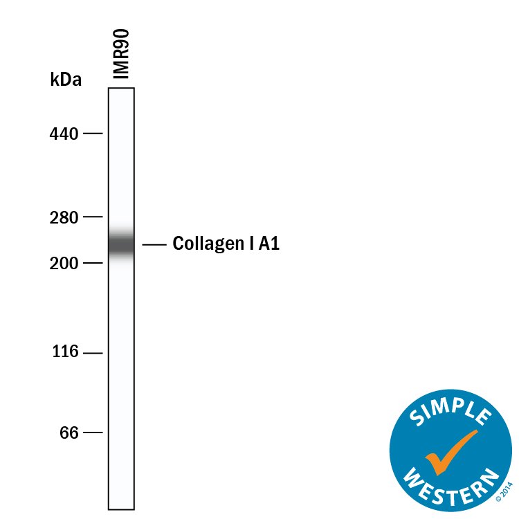
Detection of Human Collagen I alpha 1 by Simple WesternTM. Simple Western lane view shows lysates of IMR‑90 human lung fibroblast cell line, loaded at 0.2 mg/mL. A specific band was detected for Collagen I alpha 1 at approximately 232 kDa (as indicated) using 10 µg/mL of Sheep Anti-Human Pro Collagen I alpha 1 Antigen Affinity-purified Polyclonal Antibody (Catalog # AF6220) followed by 1:50 dilution of HRP-conjugated Anti-Sheep IgG Secondary Antibody (HAF016). This experiment was conducted under reducing conditions and using the 66-440 kDa separation system.
Reconstitution Calculator
Preparation and Storage
- 12 months from date of receipt, -20 to -70 °C as supplied.
- 1 month, 2 to 8 °C under sterile conditions after reconstitution.
- 6 months, -20 to -70 °C under sterile conditions after reconstitution.
Background: Collagen I alpha 1
Type I collagen is the most abundant structural protein of connective tissues such as skin, bone and tendon. It is synthesized as a procollagen molecule which is characterized by a 300 nm triple helical domain flanked by globular N- and C-terminal propeptides (1). The triple helical domain contains Gly-Xaa-Yaa triplets where Xaa and Yaa are frequently proline and hydroxyproline, respectively. The non-helical propeptides are removed by procollagen N- and C-proteinase activities so that the mature triple helices can self-assemble into collagen fibrils that provide tensile strength to tissues (1). Type I collagen is a heterotrimer that consists of two alpha 1(I) chains and one alpha 2(I) chain, although homotrimers consisting of three identical alpha 1(I) chains have also been described (2). This recombinant mini pro-alpha 1(I) collagen consists of a shortened alpha 1(I) chain with following domain structure from N- to C-terminus: N-propeptide, N‑telopeptide, the 33 most N-terminal Gly-Xaa-Yaa repeats, the 33 most C-terminal Gly-Xaa-Yaa repeats, C-telopeptide and C-propeptide. The preparation contains a mixture of the full-length molecule, pN collagen I( alpha 1) and the C-terminal propeptide. This truncated pro-alpha 1(I) collagen is a substrate for procollagen N-proteinase and procollagen C-proteinase.
- Canty, E.G. et al. (2005) J. Cell Sci. 118:1341.
- Han, S. et al. (2008) J. Mol. Biol. 383:122.
Product Datasheets
Citations for Human Pro Collagen I alpha 1 Antibody
R&D Systems personnel manually curate a database that contains references using R&D Systems products. The data collected includes not only links to publications in PubMed, but also provides information about sample types, species, and experimental conditions.
12
Citations: Showing 1 - 10
Filter your results:
Filter by:
-
Mapping of clonal lineages across developmental stages in human neural differentiation
Authors: Z You, L Wang, H He, Z Wu, X Zhang, S Xue, P Xu, Y Hong, M Xiong, W Wei, Y Chen
Cell Stem Cell, 2023-03-17;0(0):.
Species: Human
Sample Types: Organoid
Applications: IHC -
Mapping of clonal lineages across developmental stages in human neural differentiation
Authors: Z You, L Wang, H He, Z Wu, X Zhang, S Xue, P Xu, Y Hong, M Xiong, W Wei, Y Chen
Cell Stem Cell, 2023;0(0):.
Species: Human
Sample Types: Organoid
Applications: IHC -
Transcriptomic and Immunohistochemical Analysis of Progressive Keratoconus Reveal Altered WNT10A in Epithelium and Bowman's Layer
Authors: JW Foster, RN Parikh, J Wang, KS Bower, M Matthaei, S Chakravart, AS Jun, CG Eberhart, US Soiberman
Investigative Ophthalmology & Visual Science, 2021-05-03;62(6):16.
Species: Human
Sample Types: Tissue Homogenates
Applications: Western Blot -
Suppression of pancreatic ductal adenocarcinoma growth and metastasis by fibrillar collagens produced selectively by tumor cells
Authors: C Tian, Y Huang, KR Clauser, S Rickelt, AN Lau, SA Carr, MG Vander Hei, RO Hynes
Nature Communications, 2021-04-20;12(1):2328.
Species: Human
Sample Types: Tissue, Tissue Homogenates
Applications: IHC, Western Blot -
TGF-&beta2 Promotes Oxidative Stress in Human Trabecular Meshwork Cells by Selectively Enhancing NADPH Oxidase 4 Expression
Authors: VR Rao, EB Stubbs
Investigative Ophthalmology & Visual Science, 2021-04-01;62(4):4.
Species: Human
Sample Types: Cell Lysates, Whole Cells
Applications: ICC, Western Blot -
Autologous transplant therapy alleviates motor and depressive behaviors in parkinsonian monkeys
Authors: Y Tao, SC Vermilyea, M Zammit, J Lu, M Olsen, JM Metzger, L Yao, Y Chen, S Phillips, JE Holden, V Bondarenko, WF Block, TE Barnhart, N Schultz-Da, K Brunner, H Simmons, BT Christian, ME Emborg, SC Zhang
Nature Medicine, 2021-03-01;0(0):.
Species: Primate - Rhesus macaque
Sample Types: Whole Cells, Whole Tissue
Applications: ICC, IHC -
Preclinical Efficacy and Safety of a Human Embryonic Stem Cell-Derived Midbrain Dopamine Progenitor Product, MSK-DA01
Authors: J Piao, S Zabierowsk, BN Dubose, EJ Hill, M Navare, N Claros, S Rosen, K Ramnarine, C Horn, C Fredrickso, K Wong, B Safford, S Kriks, A El Maarouf, U Rutishause, C Henchcliff, Y Wang, I Riviere, S Mann, V Bermudez, S Irion, L Studer, M Tomishima, V Tabar
Cell Stem Cell, 2021-02-04;28(2):217-229.e7.
Species: Mouse
Sample Types: Whole Tissue
Applications: IHC -
Extracellular histones stimulate collagen expression in vitro and promote liver fibrogenesis in a mouse model via the TLR4-MyD88 signaling pathway
Authors: Z Wang, ZX Cheng, ST Abrams, ZQ Lin, ED Yates, Q Yu, WP Yu, PS Chen, CH Toh, GZ Wang
World Journal of Gastroenterology, 2020-12-21;26(47):7513-7527.
Species: Human
Sample Types: Cell Lysates
Applications: Western Blot -
Modeling Progressive Fibrosis with Pluripotent Stem Cells Identifies an Anti-fibrotic Small Molecule
Authors: P Vijayaraj, A Minasyan, A Durra, S Karumbayar, M Mehrabi, CJ Aros, SD Ahadome, DW Shia, K Chung, JM Sandlin, KF Darmawan, KV Bhatt, CC Manze, MK Paul, DC Wilkinson, W Yan, AT Clark, TM Rickabaugh, WD Wallace, TG Graeber, R Damoiseaux, BN Gomperts
Cell Rep, 2019-12-10;29(11):3488-3505.e9.
Species: Human
Sample Types: Cell Lysates, Whole Cells
Applications: ICC, Western Blot -
Evidence Suggesting a Role of Iron in a Mouse Model of Nephrogenic Systemic Fibrosis.
Authors: Bose C, Megyesi J, Shah S, Hiatt K, Hall K, Karaduta O, Swaminathan S
PLoS ONE, 2015-08-25;10(8):e0136563.
Species: Human
Sample Types: Cell Lysates
Applications: Western Blot -
Effect of thrombin on human amnion mesenchymal cells, mouse fetal membranes, and preterm birth.
Authors: Mogami, Haruta, Keller, Patrick, Shi, Haolin, Word, R Ann
J Biol Chem, 2014-03-20;289(19):13295-307.
Species: Human
Sample Types: Cell Lysates
Applications: Western Blot -
PDGFRbeta expression and function in fibroblasts derived from pluripotent cells is linked to DNA demethylation.
J. Cell. Sci., 2012-02-17;125(0):2276-87.
Species: Human
Sample Types: Tissue Homogenates
Applications: Western Blot
FAQs
No product specific FAQs exist for this product, however you may
View all Antibody FAQsReviews for Human Pro Collagen I alpha 1 Antibody
Average Rating: 5 (Based on 1 Review)
Have you used Human Pro Collagen I alpha 1 Antibody?
Submit a review and receive an Amazon gift card.
$25/€18/£15/$25CAN/¥75 Yuan/¥2500 Yen for a review with an image
$10/€7/£6/$10 CAD/¥70 Yuan/¥1110 Yen for a review without an image
Filter by:
