Human Rab27a Antibody Summary
Ser135-Ala218
Accession # P51159
*Small pack size (-SP) is supplied either lyophilized or as a 0.2 µm filtered solution in PBS.
Applications
Please Note: Optimal dilutions should be determined by each laboratory for each application. General Protocols are available in the Technical Information section on our website.
Scientific Data
 View Larger
Detection of Human Rab27a by Western Blot. Western blot shows lysates of human prostate tissue. PVDF membrane was probed with 2 µg/mL of Rabbit Anti-Human Rab27a Monoclonal Antibody (Catalog # MAB7245) followed by HRP-conjugated Anti-Rabbit IgG Secondary Antibody (Catalog # HAF008). A specific band was detected for Rab27a at approximately 26 kDa (as indicated). This experiment was conducted under reducing conditions and using Immunoblot Buffer Group 1.
.") View Larger
View Larger
Rab27a in SK‑Mel‑28 Human Cell Line. Rab27a was detected in immersion fixed SK-Mel-28 human malignant melanoma cell line (left panel; positive staining) and Daudi human Burkitt's lymphoma cell line (right panel; negative staining) using Rabbit Anti-Human Rab27a Monoclonal Antibody (Catalog # MAB7245) at 3 µg/mL for 3 hours at room temperature. Cells were stained using the NorthernLights™ 557-conjugated Anti-Rabbit IgG Secondary Antibody (red; Catalog # NL004) and counterstained with DAPI (blue). Specific staining was localized to cytoplasm. View our protocol for Fluorescent ICC Staining of Cells on Coverslips.
 View Larger
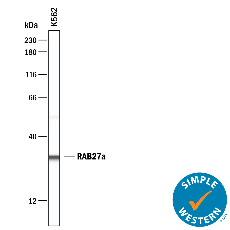
Detection of Human Rab27a by Simple WesternTM. Simple Western lane view shows lysates of K562 human chronic myelogenous leukemia cell line, loaded at 0.2 mg/mL. A specific band was detected for Rab27a at approximately 31 kDa (as indicated) using 20 µg/mL of Rabbit Anti-Human Rab27a Monoclonal Antibody (Catalog # MAB7245). This experiment was conducted under reducing conditions and using the 12-230 kDa separation system.
Reconstitution Calculator
Preparation and Storage
- 12 months from date of receipt, -20 to -70 °C as supplied.
- 1 month, 2 to 8 °C under sterile conditions after reconstitution.
- 6 months, -20 to -70 °C under sterile conditions after reconstitution.
Background: Rab27a
RAB27A (Ras-related protein Rab 27A; also GTP-binding protein Ram) is a 27-28 kDa member of the Rab27 subfamily, Rab family, Small GTPase superfamily of proteins. It is widely expressed, and found in cells diverse as mast cells, cytotoxic T cells, melanocytes, retinal pigment epithelium and pancreatic beta -cells. RAB27A plays a key role in the secretion of specialized lysosomes termed secretory lysosomes. In melanocytes, for example, RAB27A is incorporated into the melanosome membrane where it serves as a docking factor for melanophilin and myosin-Va, regulating melanosome transport to, and concentration at, sites of release. Human RAB27A is 221 amino acids (aa) in length. It contains multiple Rab family and subfamily motifs, and concludes with a C-terminal CXC prenylation sequence (aa 219‑221). There is one potential splice variant that shows a deletion of aa 146-153. Over aa 135-218, human RAB27A shares 92% and 94% aa sequence identity with mouse Rab27A and rat RAB27A, respectively.
Product Datasheets
Citation for Human Rab27a Antibody
R&D Systems personnel manually curate a database that contains references using R&D Systems products. The data collected includes not only links to publications in PubMed, but also provides information about sample types, species, and experimental conditions.
1 Citation: Showing 1 - 1
-
Neutrophil-specific interactome of ARHGAP25 reveals novel partners and regulatory insights
Authors: Sasvári, P;Pettkó-Szandtner, A;Wisniewski, É;Csépányi-Kömi, R;
Scientific reports
Species: Human
Sample Types: Protein
Applications: Western Blot
FAQs
No product specific FAQs exist for this product, however you may
View all Antibody FAQsReviews for Human Rab27a Antibody
Average Rating: 5 (Based on 1 Review)
Have you used Human Rab27a Antibody?
Submit a review and receive an Amazon gift card.
$25/€18/£15/$25CAN/¥75 Yuan/¥2500 Yen for a review with an image
$10/€7/£6/$10 CAD/¥70 Yuan/¥1110 Yen for a review without an image
Filter by:


