Human sFRP-1 Antibody Summary
Ser32-Lys314
Accession # AAB70793
Applications
Please Note: Optimal dilutions should be determined by each laboratory for each application. General Protocols are available in the Technical Information section on our website.
Scientific Data
 View Larger
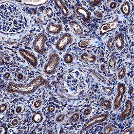
sFRP‑1 in Human Kidney. sFRP-1 was detected in immersion fixed paraffin-embedded sections of human kidney using Goat Anti-Human sFRP-1 Antigen Affinity-purified Polyclonal Antibody (Catalog # AF1384) at 1 µg/mL overnight at 4 °C. Tissue was stained using the Anti-Goat HRP-DAB Cell & Tissue Staining Kit (brown; Catalog # CTS008) and counterstained with hematoxylin (blue). Specific staining was localized to epithelial cell cytoplasm in convoluted tubules. View our protocol for Chromogenic IHC Staining of Paraffin-embedded Tissue Sections.
 View Larger
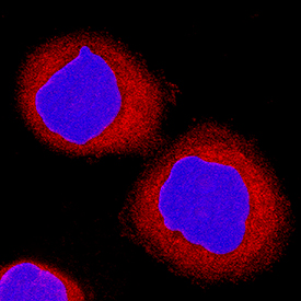
sFRP‑1 in MBA‑MB‑468 Human Cell Line. sFRP-1 was detected in immersion fixed MBA-MB-468 human breast cancer cell line using Goat Anti-Human sFRP-1 Antigen Affinity-purified Polyclonal Antibody (Catalog # AF1384) at 15 µg/mL for 3 hours at room temperature. Cells were stained using the NorthernLights™ 557-conjugated Anti-Goat IgG Secondary Antibody (red; Catalog # NL001) and counterstained with DAPI (blue). Specific staining was localized to cytoplasm. View our protocol for Fluorescent ICC Staining of Cells on Coverslips.
Reconstitution Calculator
Preparation and Storage
- 12 months from date of receipt, -20 to -70 °C as supplied.
- 1 month, 2 to 8 °C under sterile conditions after reconstitution.
- 6 months, -20 to -70 °C under sterile conditions after reconstitution.
Background: sFRP-1
Secreted Frizzled Related Proteins (sFRPs) are a family of secreted, soluble vertebrate glycoproteins which contain homology to the Wnt-binding domain of the Frizzled (Fz) family of transmembrane receptors. sFRPs are approximately 30-35 kDa in size and are comprised of 3 domains: a signal sequence; an N-terminal Fz cysteine-rich domain (CRD) with 10 conserved cysteines; and a C-terminal heparin-binding region with weak homology to Netrin. The Fz CRD mediates Wnt-binding and is present in all Fz and sFRP family members (1).
sFRP-1, also known as secreted apoptosis-related protein 2 (SARP-2), FRP and FrzA, is expressed in the embryonic kidney, eye, brain, teeth, salivary gland and small intestine, most often at sites of epithelial-mesenchyme interaction (5). Expression in the adult animal is strong in the eye, kidney, and heart and also prevalent in the brain and lung (2, 5). sFRP-1 was first characterized as a protein that enhances the sensitivity of cells to apoptotic stimuli (3) and as an antagonist of Wnt signaling in Xenopus embryos (4). It is also characterized as a tumor suppressor in breast (6) and cervical carcinomas (7). In contrast, sFRP-1 is expressed by the majority of malignant gliomas (8) and contributes to the development of uterine leiomyomas (9), suggesting that the role of sFRP-1 is dependent on cell context. sFRP-1 has diverse activities, from inducing angiogenesis (10) in a variety of in vivo models to helping regulate Wnt-4 signaling (with sFRP-2) in renal organogenesis (11). Mouse and human sFRP-1 proteins share 94% amino acid identity (1).
- Jones, S. et al. (2002) Bioessays 24:811.
- Rattner, A. et al. (1997) Proc. Natl. Acad. Sci. USA 94:2859.
- Melkonyan, H. et al. (1997) Proc. Natl. Acad. Sci. USA 94:13636.
- Finch, P. et al. (1997) Proc. Natl. Acad. Sci. USA 94:6770.
- Leimeister, C. et al. (1998) Mech. Dev. 75:29.
- Ugolini, F. et al. (2001) Oncogene 20:5810.
- Ko, J. et al. (2002) Exp. Cell. Res. 280:280.
- Roth, W. et al. (2000) Oncogene 19:4210.
- Fukuhara, K, et al. (2002) J. Clin. Endocr. Metab. 87:1729.
- Dufourcq, P. et al. (2002) Circulation 106: 3097.
- Yoshino, K. et al. (2001) Mech. Dev. 102:45.
Product Datasheets
Citations for Human sFRP-1 Antibody
R&D Systems personnel manually curate a database that contains references using R&D Systems products. The data collected includes not only links to publications in PubMed, but also provides information about sample types, species, and experimental conditions.
6
Citations: Showing 1 - 6
Filter your results:
Filter by:
-
Forced expression of Wnt antagonists sFRP1 and WIF1 sensitizes chronic myeloid leukemia cells to tyrosine kinase inhibitors
Authors: M Pehlivan, C Caliskan, Z Yuce, HO Sercan
Tumour Biol., 2017-05-01;39(5):1010428317701.
Species: Human
Sample Types: Cell Lysates
Applications: Western Blot -
Low expression of RBMS3 and SFRP1 are associated with poor prognosis in patients with gastric cancer
Am J Cancer Res, 2016-11-01;6(11):2679-2689.
Species: Human
Sample Types: Tissue Homogenates, Whole Tissue
Applications: IHC-P, Western Blot -
Blockade of Wnt signaling inhibits angiogenesis and tumor growth in hepatocellular carcinoma.
Authors: Hu J, Dong A, Fernandez-Ruiz V, Shan J, Kawa M, Martinez-Anso E, Prieto J, Qian C
Cancer Res., 2009-08-18;69(17):6951-9.
Species: Human
Sample Types: Tissue Homogenates
Applications: Western Blot -
Wnt signaling mediates experience-related regulation of synapse numbers and mossy fiber connectivities in the adult hippocampus.
Authors: Gogolla N, Galimberti I, Deguchi Y, Caroni P
Neuron, 2009-05-28;62(4):510-25.
Species: Mouse
Sample Types: Whole Tissue
Applications: IHC-Fr -
Wnt signaling antagonists are potential prognostic biomarkers for the progression of radiographic hip osteoarthritis in elderly Caucasian women.
Authors: Lane NE, Nevitt MC, Lui LY, de Leon P, Corr M
Arthritis Rheum., 2007-10-01;56(10):3319-25.
Species: Human
Sample Types: Serum
Applications: ELISA Development -
Genomics identifies medulloblastoma subgroups that are enriched for specific genetic alterations.
Authors: Thompson MC, Fuller C, Hogg TL, Dalton J, Finkelstein D, Lau CC, Chintagumpala M, Adesina A, Ashley DM, Kellie SJ, Taylor MD, Curran T, Gajjar A, Gilbertson RJ
J. Clin. Oncol., 2006-03-27;24(12):1924-31.
Species: Human
Sample Types: Whole Tissue
Applications: IHC
FAQs
No product specific FAQs exist for this product, however you may
View all Antibody FAQsReviews for Human sFRP-1 Antibody
Average Rating: 4 (Based on 2 Reviews)
Have you used Human sFRP-1 Antibody?
Submit a review and receive an Amazon gift card.
$25/€18/£15/$25CAN/¥75 Yuan/¥2500 Yen for a review with an image
$10/€7/£6/$10 CAD/¥70 Yuan/¥1110 Yen for a review without an image
Filter by:


