Human SR-AI/MSR Antibody Summary
Lys77-Leu451
Accession # P21757
Applications
Please Note: Optimal dilutions should be determined by each laboratory for each application. General Protocols are available in the Technical Information section on our website.
Scientific Data
 View Larger
Detection of Human SR‑AI/MSR by Western Blot. Western blot shows lysates of THP-1 human acute monocytic leukemia cell line untreated (-) or treated (+) with 80 nM PMA for 72 hours. PVDF membrane was probed with 1 µg/mL of Mouse Anti-Human SR-AI/MSR Monoclonal Antibody (Catalog # MAB2708) followed by HRP-conjugated Anti-Mouse IgG Secondary Antibody (Catalog # HAF018). A specific band was detected for SR-AI/MSR at approximately 80 kDa (as indicated). This experiment was conducted under reducing conditions and using Immunoblot Buffer Group 1.
 View Larger
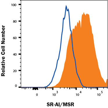
Detection of SR‑AI/MSR in M2 macrophages cells by Flow Cytometry. M2 macrophages cells treated with 50 ng/mL Recombinant Human M-CSF Protein (216-MC) for 6 days followed by an overnight polarization with 20 ng/mL Recombinant Human IL-4 Protein (204-IL) and 20 ng/mL Recombinant Human IL-13 Protein (213-ILB) were stained with Mouse Anti-Human SR‑AI/MSR Monoclonal Antibody (Catalog # MAB2708, filled histogram) or isotype control antibody (Catalog # MAB004, open histogram), followed by Phycoerythrin-conjugated Anti-Mouse IgG Secondary Antibody (Catalog # F0102B). View our protocol for Staining Membrane-associated Proteins.
Reconstitution Calculator
Preparation and Storage
- 12 months from date of receipt, -20 to -70 °C as supplied.
- 1 month, 2 to 8 °C under sterile conditions after reconstitution.
- 6 months, -20 to -70 °C under sterile conditions after reconstitution.
Background: SR-AI/MSR
The type I class A macrophage scavenger receptor (SR-AI; also MSR-AI) is a 70-80 kDa protein that belongs to the scavenger receptor superfamily (1‑3). Receptors of this family contain characteristic extracellular domains and bind to a series of generally unrelated, but negatively-charged/polyanionic ligands (1, 3). Human SR-AI is a type II transmembrane glycoprotein that is 451 amino acids (aa) in length. It contains a 50 aa cytoplasmic tail, a 26 aa transmembrane segment and a 375 aa extracellular region (4, 5). The extracellular region contains four definitive domains, with a membrane proximal spacer of 33 aa, an alpha -helical coiled-coil domain of 163 aa, a collagen-like domain of 69 aa, and a cysteine-rich C-terminus of 110 aa (4, 6). The cysteine-rich domain (CRD) forms three intrachain disulfide bonds (7). The functional form of the molecule is a 220‑230 kDa membrane-associated trimer that, in human, apparently has two disulfide bonded chains and a third noncovalently associated subunit (8, 9). Human extracellular region is 73% and 72% aa identical to bovine and mouse SR-AI extracellular region, respectively. The human gene for SR-A gives rise to three isoforms; the I isoform of 451 aa, the II isoform of 358 aa, and the III isoform of 388 aa (4, 5, 10). All are identical through the first 344 aa which includes the cytoplasmic tail through the collagenous domain. Isoform II (SR-AII) shows a severe truncation of the CRD, but is expressed on the cell surface. Isoform III (SR-AIII) has a modest truncation of the CRD, and cannot be expressed on the cell surface. However, relative to SR-AI, SR-AII is known to show differential sensitivity to LPS and receptor binding to gram‑negative bacteria (9, 11), while SR-AIII is known to be a dominant-negative isoform (10). SR-AIII may achieve this by either heterotrimerizing with SR-AI, or simply eliminating the production of SR-AI mRNA.
- Platt, N. and S. Gordon (2001) J. Clin. Invest. 108:649.
- Linton, M.F. and S. Fazio (2001) Curr. Opin. Lipidol. 12:489.
- Platt, N. and S. Gordon (1998) Chem. Biol. 5:R193.
- Matsumoto, A. et al. (1990) Proc. Natl. Acad. Sci. USA 87:9133.
- Emi, M. et al. (1993) J. Biol. Chem. 268:2120.
- Naito, M. et al. (1992) Am. J. Pathol. 141:591.
- Resnick, D. et al. (1996) J. Biol. Chem. 271:26924.
- Ashkenas, J. et al. (1993) J. Lipid Res. 34:983.
- Penman, M. et al. (1991) J. Biol. Chem. 266:23985.
- Gough, P.J. et al. (1998) J. Lipid Res. 39:531.
- Peiser, L. et al. (2000) Inf. Immun. 68:1953.
Product Datasheets
Citation for Human SR-AI/MSR Antibody
R&D Systems personnel manually curate a database that contains references using R&D Systems products. The data collected includes not only links to publications in PubMed, but also provides information about sample types, species, and experimental conditions.
1 Citation: Showing 1 - 1
-
Leptin modulates ACAT1 expression and cholesterol efflux from human macrophages.
Authors: Hongo S, Watanabe T, Arita S
Am. J. Physiol. Endocrinol. Metab., 2009-08-01;297(2):E474-82.
Species: Human
Sample Types: Cell Lysates
Applications: Western Blot
FAQs
No product specific FAQs exist for this product, however you may
View all Antibody FAQsReviews for Human SR-AI/MSR Antibody
Average Rating: 5 (Based on 1 Review)
Have you used Human SR-AI/MSR Antibody?
Submit a review and receive an Amazon gift card.
$25/€18/£15/$25CAN/¥75 Yuan/¥2500 Yen for a review with an image
$10/€7/£6/$10 CAD/¥70 Yuan/¥1110 Yen for a review without an image
Filter by:
Lysate of THP-1 human acute monocytic leukemia cell line treated with 80nM PMA for 72 hours.


