Human TCF7/TCF1 Antibody Summary
Met116-His233
Accession # P36402
Applications
Please Note: Optimal dilutions should be determined by each laboratory for each application. General Protocols are available in the Technical Information section on our website.
Scientific Data
 View Larger
Detection of Human TCF7/TCF1 by Western Blot. Western blot shows lysates of Jurkat human acute T cell leukemia cell line and MOLT-4 human acute lymphoblastic leukemia cell line. PVDF membrane was probed with 0.2 µg/mL of Goat Anti-Human TCF7/TCF1 Antigen Affinity-purified Polyclonal Antibody (Catalog # AF5596) followed by HRP-conjugated Anti-Goat IgG Secondary Antibody (Catalog # HAF019). Specific bands were detected for TCF7/TCF1 at approximately 60, 40, and 38 kDa (as indicated). This experiment was conducted under reducing conditions and using Immunoblot Buffer Group 1.
 View Larger
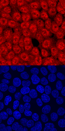
TCF7/TCF1 in HCT‑116 Human Cell Line. TCF7/TCF1 was detected in immersion fixed HCT-116 human colorectal carcinoma cell line using Human TCF7/TCF1 Antigen Affinity-purified Polyclonal Antibody (Catalog # AF5596) at 10 µg/mL for 3 hours at room temperature. Cells were stained using the NorthernLights™ 557-conjugated Anti-Goat IgG Secondary Antibody (red, upper panel; Catalog # NL001) and counterstained with DAPI (blue, lower panel). Specific staining was localized to nuclei. View our protocol for Fluorescent ICC Staining of Cells on Coverslips.
 View Larger
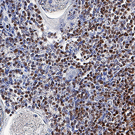
TCF7/TCF1 in Human Thymus. TCF7/TCF1 was detected in immersion fixed paraffin-embedded sections of human thymus using Goat Anti-Human TCF7/TCF1 Antigen Affinity-purified Polyclonal Antibody (Catalog # AF5596) at 3 µg/mL overnight at 4 °C. Tissue was stained using the Anti-Goat HRP-DAB Cell & Tissue Staining Kit (brown; Catalog # CTS008) and counterstained with hematoxylin (blue). Specific staining was localized to nuclei. View our protocol for Chromogenic IHC Staining of Paraffin-embedded Tissue Sections.
Reconstitution Calculator
Preparation and Storage
- 12 months from date of receipt, -20 to -70 °C as supplied.
- 1 month, 2 to 8 °C under sterile conditions after reconstitution.
- 6 months, -20 to -70 °C under sterile conditions after reconstitution.
Background: TCF7/TCF1
TCF7 (Transcription factor 7; also T cell factor 1/TCF1) is a 25‑50 kDa member of the lymphoid enhancer binding factor family of proteins with 16 isoforms. It is expressed in thymocytes and mature T cells, and serves multiple purposes. In resting cells, TCF family members are transcriptional repressors, and are 25‑32 kDa in size. Following activation, large TCF7 isoforms predominate (42‑50 kDa), and serve a transcriptional activator function. Human TCF7 is 384 amino acids (aa) in length. This is likely an activating isoform that contains a beta -catenin binding domain (aa 1‑59), a DNA-binding HMG-box (aa 269‑337), and an NLS (aa 344‑348). The use of an alternate start site at Met116 seems to characterize repressor isoforms.
Product Datasheets
FAQs
No product specific FAQs exist for this product, however you may
View all Antibody FAQsReviews for Human TCF7/TCF1 Antibody
There are currently no reviews for this product. Be the first to review Human TCF7/TCF1 Antibody and earn rewards!
Have you used Human TCF7/TCF1 Antibody?
Submit a review and receive an Amazon gift card.
$25/€18/£15/$25CAN/¥75 Yuan/¥2500 Yen for a review with an image
$10/€7/£6/$10 CAD/¥70 Yuan/¥1110 Yen for a review without an image


