Human Thioredoxin-1 Antibody Summary
Applications
Please Note: Optimal dilutions should be determined by each laboratory for each application. General Protocols are available in the Technical Information section on our website.
Scientific Data
 View Larger
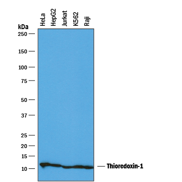
Detection of Human Thioredoxin‑1 by Western Blot. Western blot shows lysates of HeLa human cervical epithelial carcinoma cell line, HepG2 human hepatocellular carcinoma cell line, Jurkat human acute T cell leukemia cell line, K562 human chronic myelogenous leukemia cell line, and Raji human Burkitt's lymphoma cell line. PVDF membrane was probed with 0.1 µg/mL of Goat Anti-Human Thioredoxin-1 Antigen Affinity-purified Polyclonal Antibody (Catalog # AF1970) followed by HRP-conjugated Anti-Goat IgG Secondary Antibody (Catalog # HAF019). A specific band was detected for Thioredoxin-1 at approximately 12 kDa (as indicated). This experiment was conducted under reducing conditions and using Immunoblot Buffer Group 1.
 View Larger
Thioredoxin‑1 in Human Breast Cancer Tissue. Thioredoxin‑1 was detected in immersion fixed paraffin-embedded sections of human breast cancer tissue using Goat Anti-Human Thioredoxin‑1 Antigen Affinity-purified Polyclonal Antibody (Catalog # AF1970) at 15 µg/mL overnight at 4 °C. Tissue was stained using the Anti-Goat HRP-DAB Cell & Tissue Staining Kit (brown; Catalog # CTS008) and counterstained with hematoxylin (blue). View our protocol for Chromogenic IHC Staining of Paraffin-embedded Tissue Sections.
 View Larger
Thioredoxin‑1 in Human Breast Cancer Tissue. Thioredoxin‑1 was detected in immersion fixed paraffin-embedded sections of human breast cancer tissue using Goat Anti-Human Thioredoxin‑1 Antigen Affinity-purified Polyclonal Antibody (Catalog # AF1970) at 5 µg/mL overnight at 4 °C. Tissue was stained using the Anti-Goat HRP-DAB Cell & Tissue Staining Kit (brown; Catalog # CTS008) and counterstained with hematoxylin (blue). View our protocol for Chromogenic IHC Staining of Paraffin-embedded Tissue Sections.
 View Larger
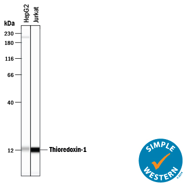
Detection of Human Thioredoxin‑1 by Simple WesternTM. Simple Western lane view shows lysates of HepG2 human hepatocellular carcinoma cell line and Jurkat human acute T cell leukemia cell line, loaded at 0.2 mg/mL. A specific band was detected for Thioredoxin-1 at approximately 12 kDa (as indicated) using 1 µg/mL of Goat Anti-Human Thioredoxin-1 Antigen Affinity-purified Polyclonal Antibody (Catalog # AF1970) followed by 1:50 dilution of HRP-conjugated Anti-Goat IgG Secondary Antibody (Catalog # HAF109). This experiment was conducted under reducing conditions and using the 12-230 kDa separation system.
Reconstitution Calculator
Preparation and Storage
- 12 months from date of receipt, -20 to -70 degreesC as supplied. 1 month, 2 to 8 degreesC under sterile conditions after reconstitution. 6 months, -20 to -70 degreesC under sterile conditions after reconstitution.
Background: Thioredoxin-1
Thioredoxins (Trxs) are a group of small ubiquitous proteins in all living cells that are key regulators of cellular redox balance (1, 2). The mammalian Trx family has three members. The Trx-1, which is a secreted and cellular protein, the mitochondria-specific Trx-2, and the Trx-like cytosolic protein p32TrxL (3-5). The active site of mammalian Trxs contains two cysteines in the conserved sequence -Y-C-G-P-C-K-. In Trx-1 the conserved cysteine residues are in positions 32 and 35, respectively. Trxs exist either in a reduced or in an oxidized state when the two cysteines at the active site form an intramolecular disulfide bridge. NADPH and the flavoprotein thioredoxin reductase can convert the oxidized Trx into the reduced Trx. Trx-1 is the only extracellular occurring thioredoxin, and is secreted by lymphocytes, hepatocytes, fibroblasts, and several tumor cells. Plasma concentrations of Trx-1 are up to 6 nM (6). In cells, Trx-1 is localized predominantly in the cytoplasm. Small amounts have been detected in the nucleus and in association with the outside surface of the cells. Expression of Trx-1 is increased under various stress conditions such as hypoxia, elevated hydrogen peroxide concentrations, photochemical oxidative stress, and viral and bacterial infections. Biological functions of Trx-1 include growth factor activity, antioxidant properties, a cofactor that provides reducing equivalents, and transcriptional regulation (1, 2). The synovial tissue of rheumatoid arthritis patients produces increased levels of Trx-1 under oxidative stress conditions, and a correlation exists between the plasma levels of Trx-1 and the severity of the disease, making Trx-1 a biomarker for this pathological condition (7, 8).
- Holmgren, A. (1985) Annu. Rev. Biochem. 54:237.
- Powis, G. and W.R. Monfort (2001) Annu. Rev. Pharm. Toxicol. 41:269.
- Deiss, L.P. and A. Kimchi (1991) Science 252:117.
- Spyrou, G. et al. (1997) J. Biol. Chem. 272:2936.
- Miranda-Vizuete, A. et al. (1998) Biochem. Biophys. Res. Commun. 243:284.
- Nakamura, H. et al. (1997) Annu. Rev. Immunol. 15:147.
- Mourice, M.M. et al. (1999) Arthritis Rheum. 42:2430.
- Jikimoto, T. et al. (2001) Mol. Immunol. 38:765.
Product Datasheets
Citations for Human Thioredoxin-1 Antibody
R&D Systems personnel manually curate a database that contains references using R&D Systems products. The data collected includes not only links to publications in PubMed, but also provides information about sample types, species, and experimental conditions.
3
Citations: Showing 1 - 3
Filter your results:
Filter by:
-
Thioredoxin increases exocytosis by denitrosylating N-ethylmaleimide-sensitive factor.
Authors: Ito T, Yamakuchi M, Lowenstein CJ
J. Biol. Chem., 2011-02-15;286(13):11179-84.
Species: Human
Sample Types: Cell Lysates
Applications: Western Blot -
Thioredoxin is required for S-nitrosation of procaspase-3 and the inhibition of apoptosis in Jurkat cells.
Authors: Mitchell DA, Morton SU, Fernhoff NB, Marletta MA
Proc. Natl. Acad. Sci. U.S.A., 2007-07-02;104(28):11609-14.
Species: Human
Sample Types: Cell Lysates
Applications: Immunoprecipitation -
Thioredoxin-related mechanisms in hyperoxic lung injury in mice.
Authors: Tipple TE, Welty SE, Rogers LK, Hansen TN, Choi YE, Kehrer JP, Smith CV
Am. J. Respir. Cell Mol. Biol., 2007-06-15;37(4):405-13.
Species: Mouse
Sample Types: Tissue Homogenates
Applications: Western Blot
FAQs
No product specific FAQs exist for this product, however you may
View all Antibody FAQsReviews for Human Thioredoxin-1 Antibody
There are currently no reviews for this product. Be the first to review Human Thioredoxin-1 Antibody and earn rewards!
Have you used Human Thioredoxin-1 Antibody?
Submit a review and receive an Amazon gift card.
$25/€18/£15/$25CAN/¥75 Yuan/¥2500 Yen for a review with an image
$10/€7/£6/$10 CAD/¥70 Yuan/¥1110 Yen for a review without an image


