Human ULBP-4/RAET1E APC-conjugated Antibody Summary
Gly30-Asp225
Accession # Q8TD07
Applications
Please Note: Optimal dilutions should be determined by each laboratory for each application. General Protocols are available in the Technical Information section on our website.
Scientific Data
 View Larger
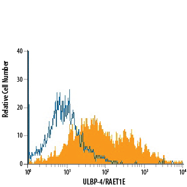
Detection of ULBP‑4/RAET1E in HepG2 Human Cell Line by Flow Cytometry. HepG2 human hepatocellular carcinoma cell line was stained with Mouse Anti-Human ULBP‑4/RAET1E APC‑conjugated Monoclonal Antibody (Catalog # FAB6285A, filled histogram) or isotype control antibody (Catalog # IC0041A, open histogram). View our protocol for Staining Membrane-associated Proteins.
Reconstitution Calculator
Preparation and Storage
- 12 months from date of receipt, 2 to 8 °C as supplied.
Background: ULBP-4/RAET1E
ULBP-4 (cytomegalovirus glycoprotein UL16 binding protein 4), also called RAET1E (retinoic acid early transcript 1E), Letal (lymphocyte effector cell toxicity activating ligand) and NKG2DL4 (NKG2D ligand 4), is a 40-50 kDa member of the ULBP/RAET1 family of cell surface proteins that function as ligands for NKG2D (1‑6). While most family members are GPI-anchored, only ULBP-4/RAET1E and ULBP-5/RAET1G express a transmembrane form (1, 4, 6, 7). Human ULBP-4 mRNA encodes 263 amino acids (aa) including a 30 aa signal sequence, a 195 aa extracellular domain (ECD), a 23 aa transmembrane domain, and a 15 aa cytoplasmic sequence. A soluble 35 kDa form diverges at aa 208 and is thought to antagonize the transmembrane form (5). Other potential splice variants of 220, 227 and 280 aa are transmembrane proteins (8). Within the ECD, ULBP-4 shares 34-41% aa sequence identity with family members (1, 7). Rodent NKG2D ligands Rae-1 alpha -epsilon are, like human ULBP and MIB proteins, distantly related to MHC class I proteins, but none of the families share significant sequence identity (2, 4). Low expression of ULBP‑4 mRNA is found in normal tissues, with high expression variably reported in the small intestine (3) and skin (4). Expression is stimulated by TNF-alpha and down‑regulated by retinoic acid (3). ULBP-4 is abnormally expressed on most colon cancer and some other tumor cell lines and virus-infected peripheral blood cells (3, 6). ULBP-4 binds and costimulates NKG2D-expressing effector cells including NK cells, NKT cells, gamma δ T cells, and CD8+ alpha beta T cells, activating cytolytic activity and/or cytokine production (3, 4, 7). In some gamma δ T cells, direct ULBP-4 binding to both TCR gamma δ and NKG2D has been demonstrated (6). ULBP-4 is also thought to function as a minor histocompatibility antigen in humans (1).
- Radosavljevic, M. et al. (2002) Genomics 79:114.
- Kondo, M. et al. (2010) Immunogenetics 62:441.
- Conejo-Garcia, J.R. et al. (2003) Cancer Biol. Ther. 2:446.
- Chalupny, N.J. et al. (2003) Biochem. Biophys. Res. Commun. 305:129.
- Cao, W. et al. (2007) J. Biol. Chem. 282:18922.
- Kong, Y. et al. (2009) Blood 114:310.
- Bacon, L. et al. (2004) J. Immunol. 173:1078.
- Cao, W. et al. (2008) Int. Immunol. 20:981.
Product Datasheets
FAQs
No product specific FAQs exist for this product, however you may
View all Antibody FAQsReviews for Human ULBP-4/RAET1E APC-conjugated Antibody
Average Rating: 4 (Based on 1 Review)
Have you used Human ULBP-4/RAET1E APC-conjugated Antibody?
Submit a review and receive an Amazon gift card.
$25/€18/£15/$25CAN/¥75 Yuan/¥2500 Yen for a review with an image
$10/€7/£6/$10 CAD/¥70 Yuan/¥1110 Yen for a review without an image
Filter by:


