Mouse B7-H3 Antibody Summary
Val29-Phe244
Accession # Q8VE98
Applications
Please Note: Optimal dilutions should be determined by each laboratory for each application. General Protocols are available in the Technical Information section on our website.
Scientific Data
 View Larger
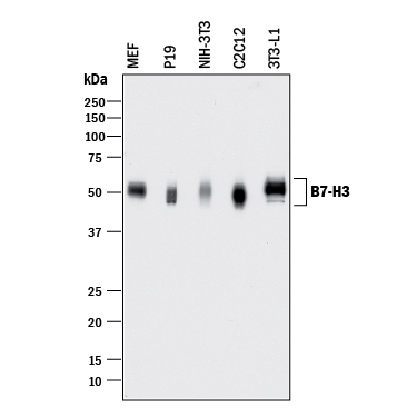
Detection of Mouse B7‑H3 by Western Blot. Western blot shows lysates of MEF mouse embryonic feeder cells, P19 mouse embryonal carcinoma cell line, NIH-3T3 mouse embryonic fibroblast cell line, C2C12 mouse myoblast cell line, and 3T3-L1 mouse embryonic fibroblast adipose-like cell line. PVDF membrane was probed with 1 µg/mL of Goat Anti-Mouse B7-H3 Antigen Affinity-purified Polyclonal Antibody (Catalog # AF1397) followed by HRP-conjugated Anti-Goat IgG Secondary Antibody (Catalog # HAF017). A specific band was detected for B7-H3 at approximately 45-55 kDa (as indicated). This experiment was conducted under reducing conditions and using Immunoblot Buffer Group 1.
 View Larger
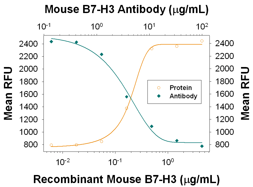
Proliferation Induced by B7‑H3 and Neutralization by Mouse B7‑H3 Antibody. Recombinant Mouse B7-H3 enhances proliferation in mouse CD3+T cells in the presence of 100 ng/mL Hamster Anti-Mouse CD3e Monoclonal Antibody (Catalog # MAB484) in a dose-dependent manner (orange line), as measured by the Resazurin (Catalog # AR002). Proliferation elicited by Recombinant Mouse B7-H3 (2 µg/mL) is neutralized (green line) by increasing concentrations of Goat Anti-Mouse B7-H3 Antigen Affinity-purified Polyclonal Antibody (Catalog # AF1397). The ND50 is typically 1.5-7.5 µg/mL.
Reconstitution Calculator
Preparation and Storage
- 12 months from date of receipt, -20 to -70 °C as supplied.
- 1 month, 2 to 8 °C under sterile conditions after reconstitution.
- 6 months, -20 to -70 °C under sterile conditions after reconstitution.
Background: B7-H3
T cells require a signal induced by the engagement of the T cell receptor and a “co‑stimulatory” signal(s) through distinct T cell surface molecules for optimal T cell expansion and activation. Members of the B7 superfamily of counter-receptors were identified by their ability to interact with co‑stimulatory molecules found on the surface of T cells. Members of the B7 superfamily include B7-1 (CD80), B7-2 (CD86), B7‑H1 (PD-L1), B7-H2 (B7RP-1), B7-H3, and PD-L2 (1). B7-H3 is expressed at very high levels in immature dendritic cells at moderate levels on mature dendritic cells, LPS stimulated immature dendritic cells and LPS stimulated monocytes, and at low levels on resting monocytes. B7-H3 binds to activated T cells via an as-of-yet identified receptor. B7-H3 co-stimulates proliferation of T cells and interferon-gamma (IFN-gamma ) production and enhances the induction of cytotoxic T cells. B7-H3 shares 20‑27% amino acid (aa) identity with other B7 family members (2). Murine B7-H3 is a 259 aa protein containing an extracellular domain, a transmembrane domain and a cytoplasmic domain. Mouse and human B7-H3 share 87% aa identity (3).
- Coyle, A.J. and J.-C. Gutierrez-Ramos (2001) Nature Immunol. 2:203.
- Chapoval, A.I. et al. (2001) Nature Immunol. 2:269.
- Sun, M. et al. (2002) J. Immunol. 168:6294.
Product Datasheets
Citations for Mouse B7-H3 Antibody
R&D Systems personnel manually curate a database that contains references using R&D Systems products. The data collected includes not only links to publications in PubMed, but also provides information about sample types, species, and experimental conditions.
2
Citations: Showing 1 - 2
Filter your results:
Filter by:
-
The immune checkpoint B7-H3 (CD276) regulates adipocyte progenitor metabolism and obesity development
Authors: E Picarda, PM Galbo, H Zong, MR Rajan, V Wallenius, D Zheng, E Börgeson, R Singh, J Pessin, X Zang
Science Advances, 2022-04-27;8(17):eabm7012.
Species: Mouse
Sample Types: Cell Lysates
Applications: Western Blot -
Origination of new immunological functions in the costimulatory molecule B7-H3: the role of exon duplication in evolution of the immune system.
Authors: Sun J, Fu F, Gu W, Yan R, Zhang G, Shen Z, Zhou Y, Wang H, Shen B, Zhang X
PLoS ONE, 2011-09-13;6(9):e24751.
Species: Mouse
Sample Types: Cell Lysates
Applications: Western Blot
FAQs
No product specific FAQs exist for this product, however you may
View all Antibody FAQsReviews for Mouse B7-H3 Antibody
There are currently no reviews for this product. Be the first to review Mouse B7-H3 Antibody and earn rewards!
Have you used Mouse B7-H3 Antibody?
Submit a review and receive an Amazon gift card.
$25/€18/£15/$25CAN/¥75 Yuan/¥2500 Yen for a review with an image
$10/€7/£6/$10 CAD/¥70 Yuan/¥1110 Yen for a review without an image

